Medical devices
search
news
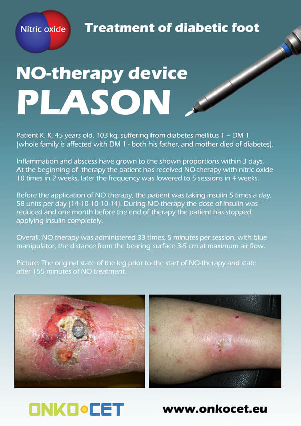
The PDF with the short report with pictures from the therapy of a diabetic foot can be viewed or downloaded here.
The pictures from the treatment of unhealing wounds an be found here:
http://www.onkocet.eu/en/produkty-detail/220/1/
The pictures from the treatment of unhealing wounds an be found here:
http://www.onkocet.eu/en/produkty-detail/293/1/

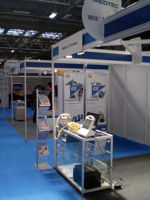
ONKOCET Ltd. has exhibited the devices from its portfolio on the MEDTEC UK exhibition in Birmingham, April 2011 through our partner Medical & Partners.
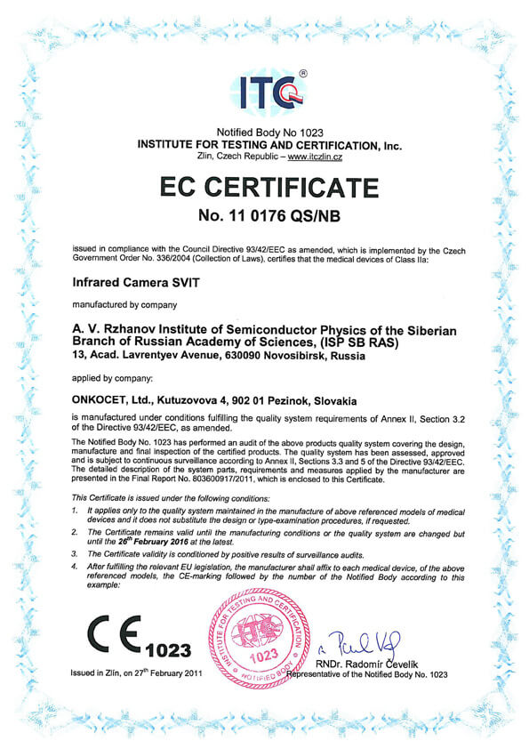 The ONKOCET company has successfully reached the certification of yet another medical device, Infrared Camera SVIT. The Certificate can be found here. The videos from the device operation can be found here.
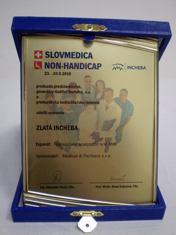 Our device, the non-invasive blood analyzer AMP has won the Golden Incheba prize at a medical exhibition SLOVMEDICA - NON-HANDICAP 2010. A big thank you goes to the organizers of the exhibition for acknowledging the quality of our device and to the exhibitor, the Medical & Partners company, for introduction of the AMP device to the medical public again.
We are pleased to inform our business partners, that our company has succesfully finished the certification process of Concor Soft Contact Lenses.
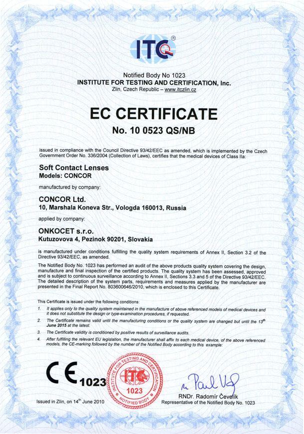 You can find the certificate here.
More information on Concor Soft Contact Lenses go to section Medical preparations/Concor soft contact lenses, or follow this link.
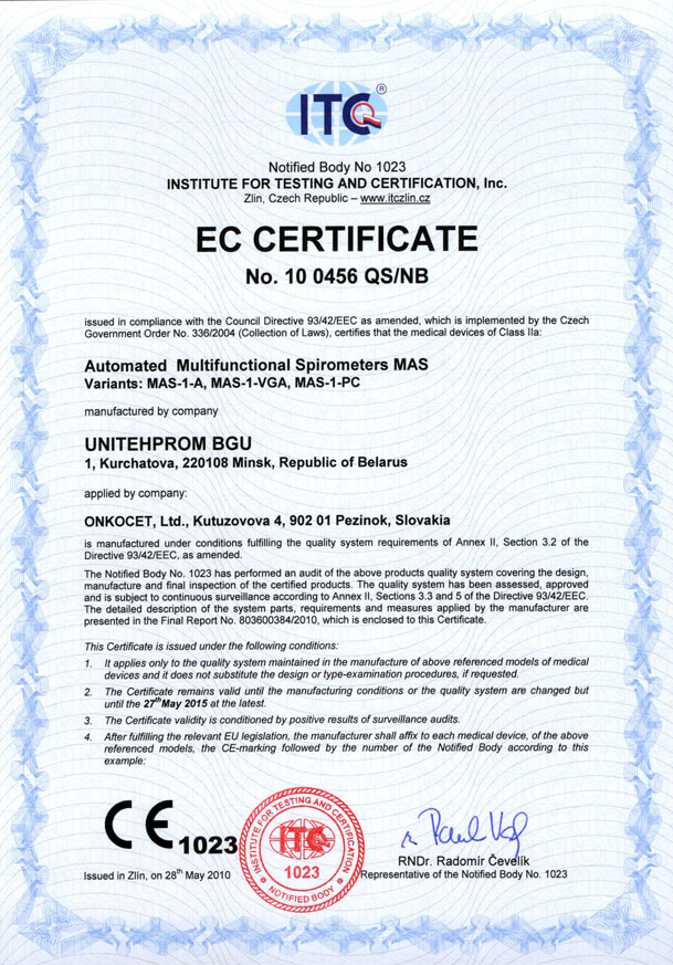 Our company has finished the certification process for another medical device, computerized spirometer MAS-1K with oximeter. You can find the device certificate here.
.jpg) Since May 2010 there is a new version of AMP device available.
Since May 2010 there is a new version of AMP device available.Follow this link if you want to see the pictures and specifications of the device.
http://www.onkocet.eu/en/produkty-detail/293/1/
 Dear partners,
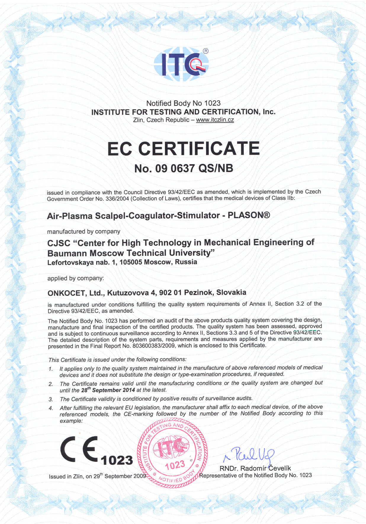
 In October 2009 we have received CE certificate for another device from our portfolio, NO therapeutical device PLASON. You can find more information about this revolutionary device, used for healing of unhealing wounds, diabetic foot, or for cosmetical purposes, at our webpage, section "Medical devices" -> PLASON-NO Therapy.
.gif)
Best regards
Team of ONKOCET Ltd. company
Introduction
Infrared Camera SVIT
Please see the video presentations of the SVIT Infra-red camera in use for visual information on how IR camera works
![]()
![]()
In the region of medicine thermal-vision camera brings the possibility to carry out the thermograph inspection of patients for the purpose of the early, preventive diagnosis of a whole series of diseases before the appearance of complaints of patient and destructive changes in the cloths. Basic merit of thermal-vision diagnostics is its absolute harmlessness and non-invasive action. With the aid of the thermal-vision camera it is possible to conduct repeated measurements of the individual sections of the skins of patient with the accumulation of information about the state of organism in the medical database. Thermal imaging system SVIT possesses the temperature sensitivity record in comparison with the classical medical thermal imaging systems: SD (standard deviation) of noise in the regime of the real functioning of instrument on the majority of matrix elements corresponds to temperature of approximately 0,025 °C. High temperature permission is especially important with the use of an instrument in medicine, since it allows recording of the low contrasting sections of thermograms, important from the diagnostic point of view. Thermal imaging system with the temperature permission up to 0,1 °C leads to smearing of low-contrast centers on the thermograms, and the elements of fine structure (vascular figure, fine focal hyper- and hypothermia) become invisible, being converted into the large spots. In this case the increased spatial resolution of such thermal-vision cameras (256x256 of elements in the sequence) is simply superfluous.
The main divisions of medicines, in which thermal-vision diagnostics of patients is most effective:
· the screening inspection of the workers of industrial enterprises, companies and so forth
· experimental medicine (study of new medicinal forms and functional physiological loads influence) on the human organism
· mammalogy (study of the mammary glands of women for the purpose of conducting preventive measures and development of tumor new formations)
· clinical diagnostics of inflammatory processes (rheumatoid arthritis, primary deformations, osteoarthrosis, arm-blade periarthritis, vibration sickness, polyneuropathy, sacroiliitis, spondylarthritis, the defeat of spine, the inflammatory processes of gall bladder, thyroid gland and others)
· oncology (early stages and differential diagnostics)
· traumatology [burns, freezings (state of vessels) and of other with the subsequent control of the effectiveness of the treatment of injuries, defeat of nerves, breaks]
· angiology (diagnostics of phlebitis and the varicose expansions of veins, diabetic angiopathy)
· fast diagnosis of the general hyperthermy of the open sections of the body of a human (atypical pneumonia, the fever of different etiology). At present, the thermal-vision method of diagnostics of atypical pneumonia is used effectively at airports and places of the mass accumulation of people)
· rapid diagnosis of LOR diseases (maxillary sinusitis, frontal sinusitis, sinusitis), thermal-vision control in sport medicine, physiotherapy, cosmetology.
Thermal-vision camera SVIT is intended for the use in medicine, science and industry for the forming of the thermal image of object (thermogram) and measurement of the temperature at any point of object without the physical contact with it. High temperature permission and the frame frequency of camera allow obtaining clear high contrast thermograms of objects in the regime of real time. This provides the possibility of the effective instrument usage in different regions of science, technology, medicine.
Thermal imaging system SVIT is the thermograph camera of the third generation, which works in real time. As photoreceiver element serves the focal matrix of semiconductor capacitors on the basis of indium arsenide (InAs) (Fig. 1). Camera is intended for temperature measuring and analysis of the static and live pictures of the thermal condition of objects. Stationary camera showed good results in the solution of the problems of medical diagnostics of the diseases by the methods of medical thermography, and also during the solution of number of technical and scientific problems in different branches of national economy, for example, for the measurement of the dynamic thermal images of the surface of the rotary furnaces with the production of cement.
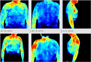
Photosensitive matrix records infrared radiation of any hot bodies, including the intrinsic emission of human skin.
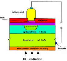
Fig. 1 The structure of the photon receiver
All material bodies with the temperature above -273 °C (0 °K), emit electromagnetic radiation, which in accordance with the Planck formula can be presented in the form, shown to Fig.2 (dependence of the emission of photons on the wavelength at two temperatures of blackbody is shown). With an increase in the temperature of object the number of the emitted quanta of radiation - (IR-radiation) with the fixed wavelengths increases. The emitted light quanta, including invisible (infrared) with a wavelength of > 1 mkm can be registered by the sensors of infrared radiations (semiconductor photon sensors).
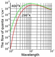
Fig.2 Dependence of the flow density of quanta, emitted by blackbody at two temperatures, on the wavelength.
The spectral range of the proposed matrix is 2,65-3,05 mkm. From the point of view of developers, for solving the number of thermograph problems, this is very convenient spectral range. This, connected with the fact that for the photon receivers the relation of the number of information falling quanta of radiation (for example for T=30 °C, certain temperature of the surface of the skin of man) and parasitic background quanta (temperature of the surrounding background of t=25 °C) increases with the decrease of the wavelength of incident radiation. So for the spectral range 8,5-12 mkm, characteristic for the receivers on the basis of compounds mercury-cadmium-tellurium relation composes 1,08, for the range 7,5-8 mkm (receiver on the basis of the superlattices of AlGaAs/GaAs) - 1,1, for the range 4,5-5 mkm (receivers on the basis of InSb and silicide of platinum) - 1,13, for range 3,5-3,9 (receivers on the basis of InSb and silicide of platinum) - 1,23, for the range 2,65-3,05 mkm (matrix on the basis of InAs of matrix) - 1,3 and for the range 1,4-1,8 mkm - 1,6. As a whole this gives the possibility to matrices in the short-wave region to more easily record small temperature contrasts with the objects. Furthermore, with the decrease of the wavelength of incident radiation decreases the parasitic flow of room background, which simplifies the reading circuits of signals.
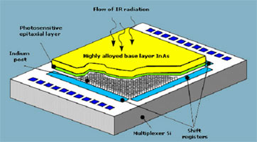
Fig.3 Structure of the hybrid microcircuit
The elements of focal matrix convert light quanta into the electric charges, which are read by the silicic multiplexer (Fig.3), are strengthened, they are preliminarily processed by electronic circuit and are transferred to the computer (Fig.4). On the screen of monitor we obtain the thermal-vision image of object (thermogram) as a result.
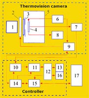
Fig.4 Block diagram of thermal-vision camera.
1 - objective, 2 - the device of calibration, 3 - cold diaphragm, 4 - matrix [FPU], 5 - vacuum cryostat with the clear window, 6 - generator of the managers of pulse and constant stresses, 7 - amplifier with the differential output, 8 - temperature gauge [FPU] the aic weapon of the switching on of the bias voltage of the base layer of InAs, 9,14 - units for control and synchronization, 10 - ACP, 11 - adder, 12 - dispatcher of memory, 13,16 - memory banks, 15 - coupling unit with the personal computer, 17 - the personal computer
In technical sense one of the advantages of the thermal imaging system of " SVIT" appears to be the fact that this thermal imaging system is built on the basis of matrix IR- detector. This advantage is manifested in the comparison with the thermal imaging systems, which use the internal scanning systems, and are currently available at the world market. In connection with the usage of the building-up principle of information signal matrix thermal imaging systems, other conditions being equal, win in the scanning systems on the basis of such parameters as reliability, sensitivity, speed and the spatial resolution.
Thermal imaging system of "SVIT" possesses the temperature sensitivity record in comparison with the classical medical thermal imaging systems: SD (standard deviation) of noise in the regime of the real functioning of instrument on the majority of matrix elements corresponds to temperature of approximately 0,025 °C. High temperature permission is especially important concerning the usage of an instrument in medicine, since it allows recording of weakly contrasting sections of thermograms important from the diagnostic point of view. Thermal imaging system with the temperature permission of 0,1 °C leads to smearing of low-contrast centers on the thermograms, and the elements of fine structure (vascular figure, fine focal hyper- and hypothermia) become invisible, being converted into the large spots. In this case the increased spatial resolution of such thermal-vision cameras (256x256 of elements in the sequence) is simply superfluous.
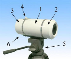
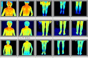
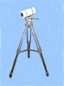
Fig. 5 The common form of thermal-vision camera in the collection.
1 - section of cryostat with the cooled focal matrix,
2 - detachable objective and the unit of calibration,
3 - detachable electronics,
4 - neck for the filling of liquid nitrogen,
5 - camera stand
6 - arrangement of joint under the standard high-speed cable USB of 2.0 A/B of cable (DUB-C5AB).