Medical devices
search
news
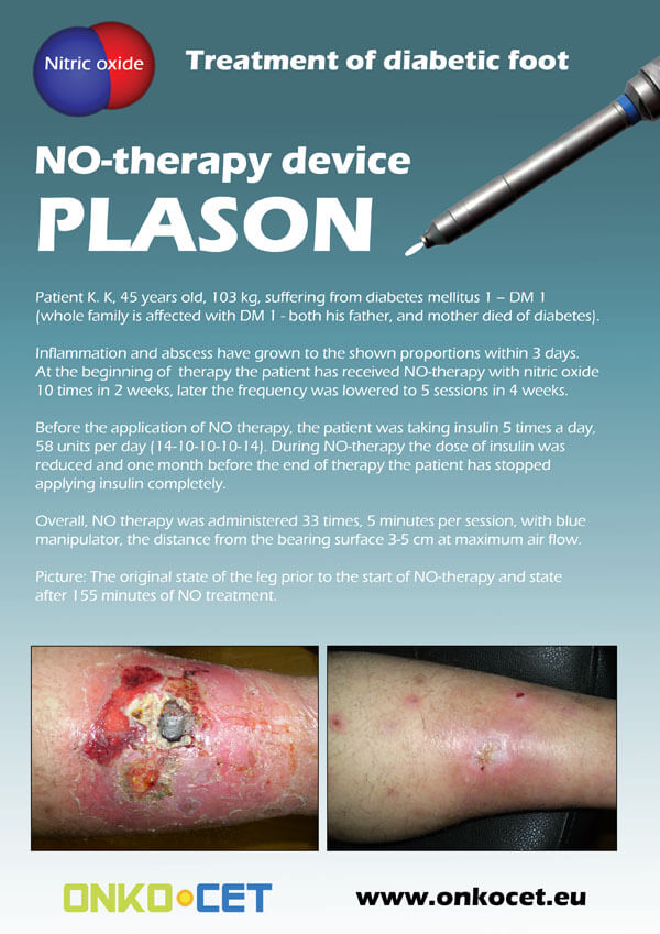
The PDF with the short report with pictures from the therapy of a diabetic foot can be viewed or downloaded here.
The pictures from the treatment of unhealing wounds an be found here:
http://www.onkocet.eu/en/produkty-detail/220/1/
The pictures from the treatment of unhealing wounds an be found here:
http://www.onkocet.eu/en/produkty-detail/293/1/
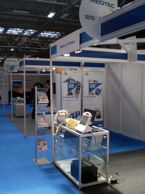
ONKOCET Ltd. has exhibited the devices from its portfolio on the MEDTEC UK exhibition in Birmingham, April 2011 through our partner Medical & Partners.
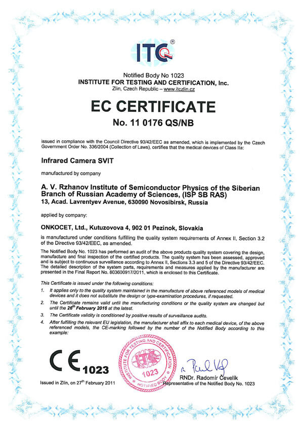 The ONKOCET company has successfully reached the certification of yet another medical device, Infrared Camera SVIT. The Certificate can be found here. The videos from the device operation can be found here.
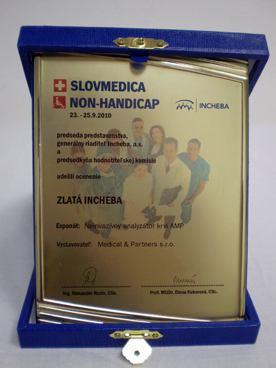 Our device, the non-invasive blood analyzer AMP has won the Golden Incheba prize at a medical exhibition SLOVMEDICA - NON-HANDICAP 2010. A big thank you goes to the organizers of the exhibition for acknowledging the quality of our device and to the exhibitor, the Medical & Partners company, for introduction of the AMP device to the medical public again.
We are pleased to inform our business partners, that our company has succesfully finished the certification process of Concor Soft Contact Lenses.
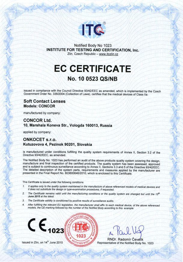 You can find the certificate here.
More information on Concor Soft Contact Lenses go to section Medical preparations/Concor soft contact lenses, or follow this link.
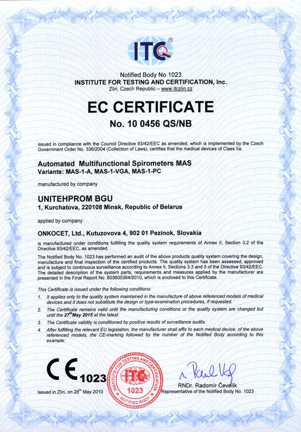 Our company has finished the certification process for another medical device, computerized spirometer MAS-1K with oximeter. You can find the device certificate here.
.jpg) Since May 2010 there is a new version of AMP device available.
Since May 2010 there is a new version of AMP device available.Follow this link if you want to see the pictures and specifications of the device.
http://www.onkocet.eu/en/produkty-detail/293/1/
 Dear partners,
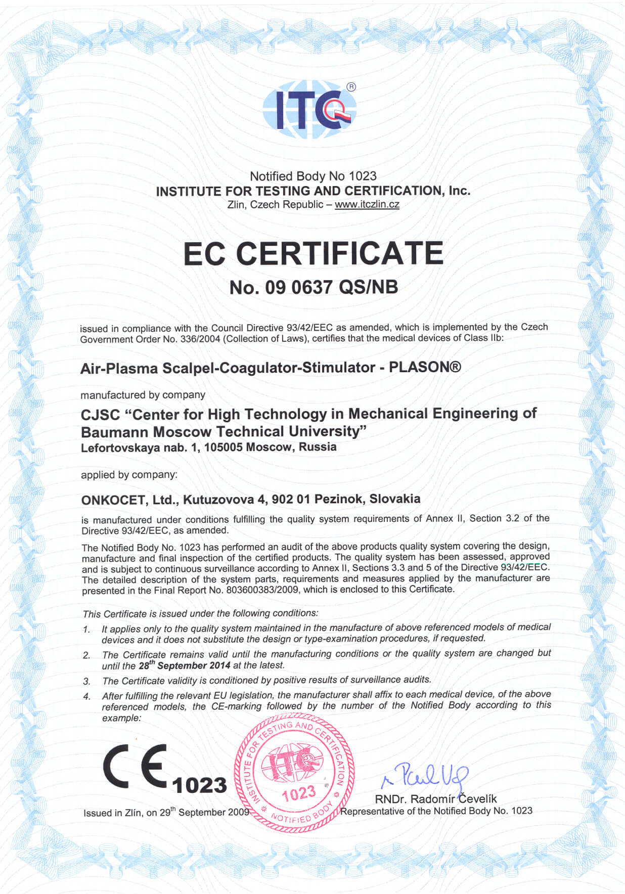
 In October 2009 we have received CE certificate for another device from our portfolio, NO therapeutical device PLASON. You can find more information about this revolutionary device, used for healing of unhealing wounds, diabetic foot, or for cosmetical purposes, at our webpage, section "Medical devices" -> PLASON-NO Therapy.
.gif)
Best regards
Team of ONKOCET Ltd. company
Assessment of mammary gland EI images
Procedure for assessment of the mammary gland electrical impedance images.
M. Korotkova (1), A. Karpov (2)
(1) Clinical Hospital Nr.9, Yaroslavl, Russia
(2) SIM-Technika, Russia.
Abstract- We analyzed more than 2000 electroimpedance images of the mammary gland in norm and with pathology. The research was carried out utilizing the electrical impedance computer tomograph «MEIK»® (current 0.5 mA, frequency 50 kHz). The following particularities of the electrical impedance image were established. In the norm the electrical impedance image reflected a regular anatomic structure of a mammary gland and the index of the mean electrical conductivity corre-sponded to the age norm. In case of pathology either changes of the anatomic structure of a mammary gland or changes of its mean electrical conductivity index were observed. Thus, estimation of the electroimpedance image should include both a visual analysis of the image as well as quantitative. The vis-ual estimation of the electroimpedance image includes analysis of the following details: the mammary gland contour (presence of deformations, infiltration); the mammary gland anatomy (changes of anatomy, displacement of internal structures, peri-focal infiltration); the lacteal sinus zone (visualization, dilata-tion); local changes of electrical conductivity (presence of hypo- or hyperimpedance areas). The quantitative analysis includes the following particulars: an index of mean electrical conductivity and a histogram of electrical conductivity distri-bution; comparison of histograms with a norm; an index of mean electrical conductivity of the area in question. The article is illustrated with electroimpedance mammograms and tables...
Procedure for assessment of the mammary gland electrical impedance images.pdf
PDF File: 400 kB

