Medical devices
search
news
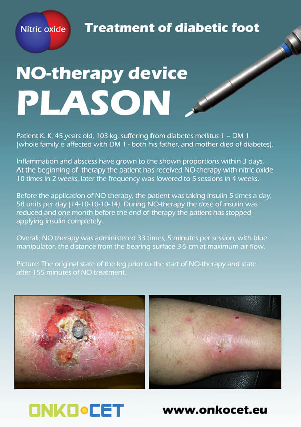
The PDF with the short report with pictures from the therapy of a diabetic foot can be viewed or downloaded here.
The pictures from the treatment of unhealing wounds an be found here:
http://www.onkocet.eu/en/produkty-detail/220/1/
The pictures from the treatment of unhealing wounds an be found here:
http://www.onkocet.eu/en/produkty-detail/293/1/
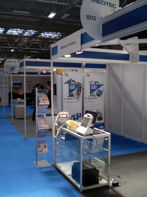
ONKOCET Ltd. has exhibited the devices from its portfolio on the MEDTEC UK exhibition in Birmingham, April 2011 through our partner Medical & Partners.
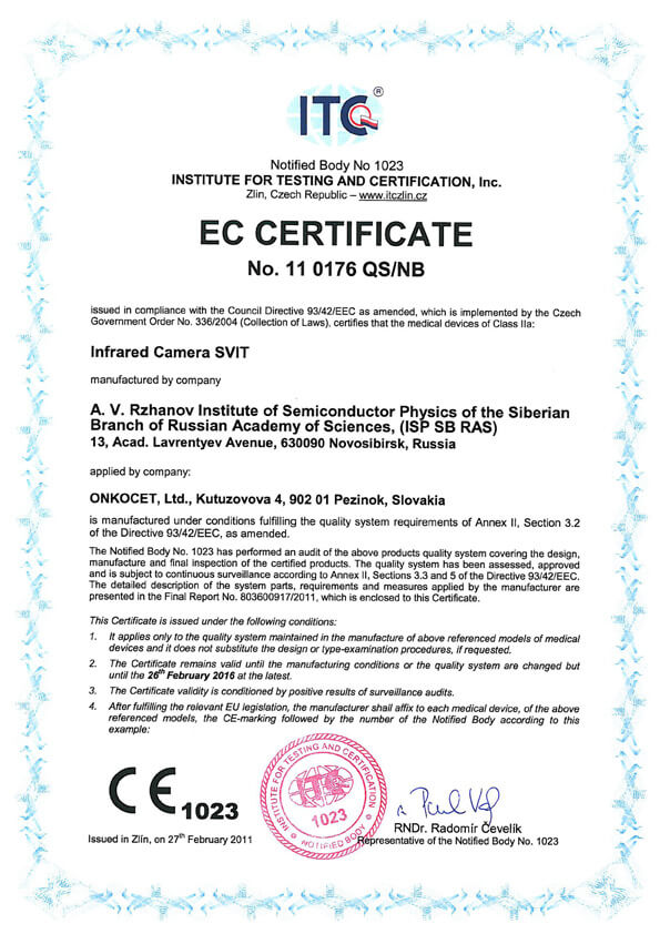 The ONKOCET company has successfully reached the certification of yet another medical device, Infrared Camera SVIT. The Certificate can be found here. The videos from the device operation can be found here.
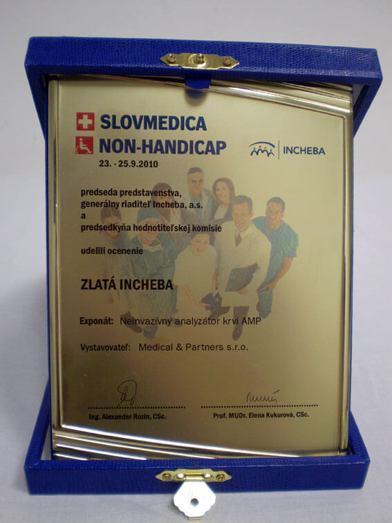 Our device, the non-invasive blood analyzer AMP has won the Golden Incheba prize at a medical exhibition SLOVMEDICA - NON-HANDICAP 2010. A big thank you goes to the organizers of the exhibition for acknowledging the quality of our device and to the exhibitor, the Medical & Partners company, for introduction of the AMP device to the medical public again.
We are pleased to inform our business partners, that our company has succesfully finished the certification process of Concor Soft Contact Lenses.
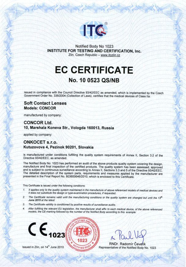 You can find the certificate here.
More information on Concor Soft Contact Lenses go to section Medical preparations/Concor soft contact lenses, or follow this link.
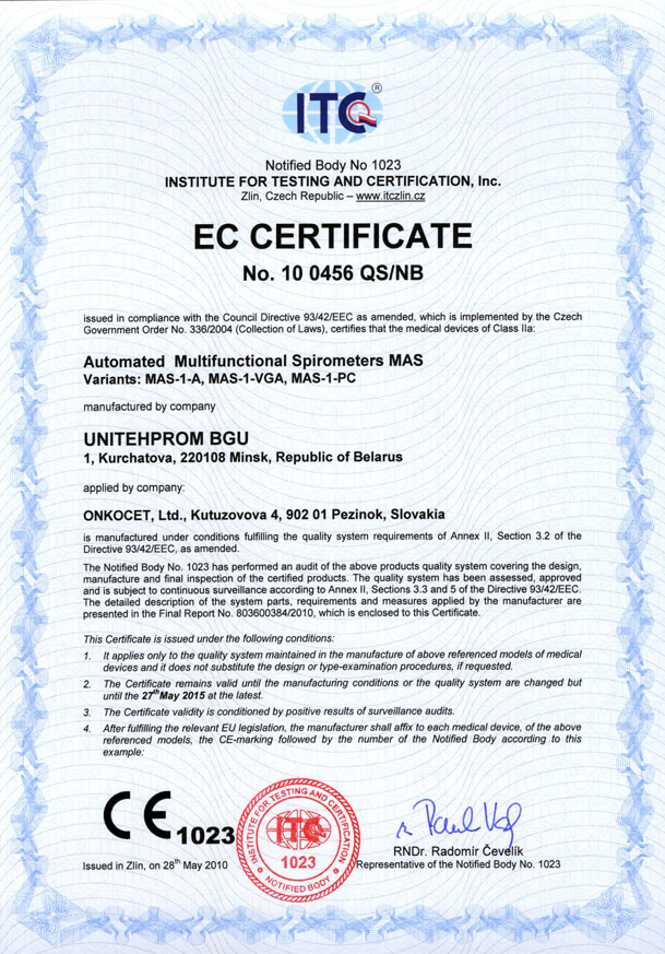 Our company has finished the certification process for another medical device, computerized spirometer MAS-1K with oximeter. You can find the device certificate here.
.jpg) Since May 2010 there is a new version of AMP device available.
Since May 2010 there is a new version of AMP device available.Follow this link if you want to see the pictures and specifications of the device.
http://www.onkocet.eu/en/produkty-detail/293/1/
 Dear partners,
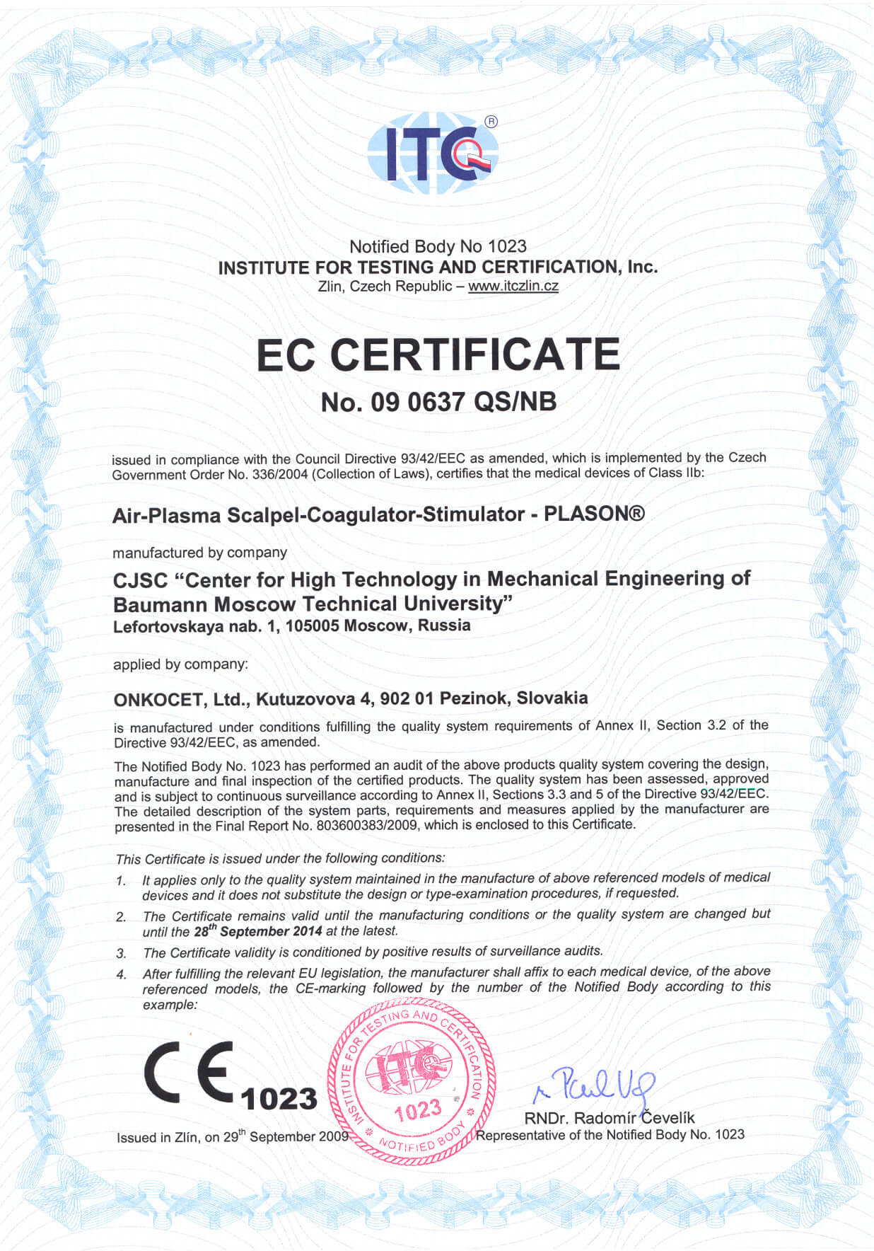
 In October 2009 we have received CE certificate for another device from our portfolio, NO therapeutical device PLASON. You can find more information about this revolutionary device, used for healing of unhealing wounds, diabetic foot, or for cosmetical purposes, at our webpage, section "Medical devices" -> PLASON-NO Therapy.
.gif)
Best regards
Team of ONKOCET Ltd. company
Description of some of the examined parameters

Description of some of the 130 examined parameters of ANESA measurement
Description of metabolic and biochemical indices, covered by Malykhin-Pulavskiy USPIH Software
Electrolyte metabolism:
10. Calcium is a cation, constituting a part of blood cells and electrolytes. Its normal concentration in the blood plasma ranges from 2.02 to 2.2 mmol/l. It is regulated by parathyroid gland, bone tissue and pituitary thyrotropic hormone. The regulation of calcium in the organism is attained by triiodothyronine and tetraiodothyronine metabolism optimization, as well as by creatinine kinase of the muscles and heart.
11. Magnesium is a cation, constituting a part of blood cells. Its primary functional purpose is participation in the development of conductivity and contractility of those muscles, constituting a part of vascular wall. Magnesium normal value ranges from 0.7 to 1.0 mmol/l. It is regulated by the activation of creatinine phosphatase phosphorylation. In case magnesium level is less than 0.7 mmol/l, clinical picture of the disease is characterized by neuromuscular hyperexcitability, spasmophilia, and asthenic feelings.
12. Potassium is a cation, being presented in the blood plasma in the concentration 4.14 - 4.56 mmol/l. Its primary functional purpose is to participate in the neuromuscular conductivity. The gastrointestinal tract and kidney play a significant role in the regulation of potassium metabolism. Hypokalemia threatens with severe consequences. It may be observed in Conn's syndrome and can accompany occasional myoparalysis, combined with migraines, epilepsy, and progressive muscular dystrophy. Hypokalemia can appear as a result of potassium loss through the gastrointestinal tract and kidneys, and due to the diabetic acidosis. Hyperkalemia is associated with the cardiac and renal malfunctions.
13. Sodium is a primary cation in the blood plasma. Its normal value is 130.5 - 156.6 mmol/l. Changes in the concentration relate to the changes in plasma specific conductivity, which normally makes up 0.72 +- 1 Ohm/cm. Changes to the regulation of sodium in the organism are associated with kidney diseases, gastrointestinal disturbances, as well as with the impairment of contractility. Rise in venous blood pressure causes hydrostatic pressure to increase. When hydrostatic pressure is higher than oncotic pressure, this can cause electrolytes out of intercellular space. This phenomenon leads to hypovolemia and activates juxtaglomerular apparatus of kidneys. This results in stimulation of the adrenal cortex and increase in aldosterone secretion. In a whole, these factor combinations give rise to the organotrophic disorder. All these factors bring on changes in the diurnal diuresis.
The system of blood fibrillation:
14,15,16,17 The most important factors of this system are plasma kinins. Practically, they form kinin system, which supports regulation of the local and general blood circulation, as well as vascular permeability. The main mechanisms are interactions between plasma kallikreins and tissue kallekreins (of pancreas, salivary glands, kidneys and intestinal wall). As the final result, this interaction leads to the beginning and to the end of blood fibrillation. It is observed that there should be an interval, exceeding 30 seconds, between the beginning and the end of blood fibrillation. The platelets (containing arachidonic acid) play an important role in the formation of this time interval. Haematocrite is of considerable importance in the formation of interval between the beginning and the end of blood fibrillation.
The fermentative system:
18,19,20,21,22 AST and ALT are enzymes catalyzing intermolecular amino group transfer between amino acids and keto acids.
As a result of the interaction between these transferases, oxaloacetic, pyruvic and glutamic acids. Elevations of aminotransferase activity, especially of AST, are observed in the myocardial diseases. Elevations in ALT activity is observed in viral hepatitis type A. Besides, elevation of ALT activity is seen with acute myocardial infarction, but this elevation is not so sharp, as compared to AST activity. Simultaneous determination of these two aminotransferases is a very valuable diagnostic test. In health, AST-ALT activity ratio (de Ritis coefficient) equals to 1.33+- 0,42. This coefficient reduces in the patients with viral hepatitis and rises in the patients with acute myocardial infarction.
23. Amylase is an enzyme that breaks carbohydrates (starch etc.) down into glucose. It facilitates glucose uptake from the blood. It is secreted with saliva into oral cavity, where it begins to break starch down, or with pancreatic juice into duodenum. Acid gastric juice inactivates amylase. The experiments have shown that after 80 g glucose was administered, amylase administration ensured that blood sugar indices remained normal. 86% of patients with diabetes have a small amount of amylase in their intestinal secretion. Being an enzyme produced by pancreas, amylase level in the blood and urine rises sharply, in the progress of pancreatitis
24. Bilirubin is a pigment, formed as a result of hemoglobin breakdown, and to a lesser extent as a result of megaglobin breakdown.
The determination of total bilirubin and its fractions is very important for the differential diagnostics of jaundice of various etiology. The elevation of unconjugated bilirubin level in the blood and tissues is determined with hemolytic jaundice. In hepatocellular jaundice, hepatocytes are being destructed , conjugated bilirubin excretion into the bile capillaries is impaired and it leaks into blood, where its level elevates significantly. Moreover, ability of the hepatocytes to synthesize bilirubin-glucuronides reduces. Consequently, unconjugated bilirubin level elevates. In obstructive jaundice, the biliary excretion is impaired, which leads to steep elevation of conjugated bilirubin level.
27. The concentration of plasma protein. Plasma proteins are divided into three groups: albumins, globulins and fibrinogen. Being colloids, proteins bind and hold water, preventing its outflow from the bloodstream. Some plasma proteins, fibrinogen in particular, are basic components of blood clotting. Blood plasma proteins are one of the most important circulatory buffers, which support level of blood cations by forming nondialyzable compounds with them. The clinical practice has evidenced conditions, defined by the change of total plasma proteins. Hyperproteinemia is a increase of total protein concentration in the plasma. It is seen with diarrhea, vomiting, obstruction of the upper small intestine and water loss. Hypoproteinemia, i.e. a decrease in the total protein concentration, is seen in the patients with neurotic syndrome. In addition, it occurs with hepatocytic lesions, and with the disorder of renal filter (lipoid nephrosis). Generally, we may relate hyperproteinemia to hyperglobulinemia and hypoproteinemia to hypoalbuminemia.
Oxygen consumption and transportation:
28. Plasma density. The plasma density is defined by total amount of plasma cations and anions. 1048 - 1055 g/cm3 is a norm. Changes in the plasma density are attributed to the disorders of water metabolism. These processes are greatly effected by antidiuretic hormone. It can be seen with Conn's syndrome, which is associated with the changes in aldosterone level. When plasma density is lower 1056, this results in blood pressure instability, hypodynamia, sometimes convulsions.
29. Circulatory blood volume. The volume of circulatory blood is a genetic value and makes up 68-70 ml/kg for men and 65-69 ml/kg for women. The changes of these values are attributed to water and electrolyte imbalance, as well as to the diseases of intestinal tract and kidneys.
30. The minute volume of circulatory blood. This is a value resulting from the functional state of organism, and is connected with respiratory rate and heart rate. The average values, calculated for a person weighing 70 kg, are 4-4.5 ml/min.
31. Oxygenation rate. It is a rate of oxidative processes, taking place in erythrocyte and in the cells. This value is formed under the influence of lipid peroxidation, which defines permeability of cell membranes, composed of lipoprotein complexes. Also, this value is related to the state of liver, gastrointestinal tract, and kidneys. The oxygenation rate is affected significantly by the temporal relations of blood circulation in the systemic and pulmonary circulation.
32. Surface of gaseous exchange. It is a erythrocyte respiratory surface, which averages 350000 cm2.- 43000 cm2. Value of the surface of gaseous exchange varies with the corpuscular volume, age, weight and sex.
33. Vital lung capacity. This is a value, representing lung ability to receive blood circulation minute volume, which defines alveolar surface area, participating in respiration.
34. Transportation of oxygen. It is a value, depending on the functional and morphological state of systemic and pulmonary circulation, and first of all on: lungs, heart, liver and gastrointestinal tract.
35. Quantity of consumed oxygen per 100 gram of brain tissue is associated with a complex cause, involved in the redox processes, lipid peroxidation reactions, and with a state of thyroid blood circulation regulation. Thyroid blood circulation determines the transportation of oxygen and its consumption by inner organs due to the activation of thyroid hormones T3 and T4.
This index depends on the activation or inactivation of organ oxygen consumption. This value averages 2.5-3.5 ml/100 g tissue for adults and 3.5-6 ml/100 g tissue for children.
36. Arterial blood oxygenation is related to hemoglobin ability to bind oxygen and oxygenate tissues. To a great extent, this process is affected by thyroxin. Thyroxin splits oxidation and phosphorylation processes, reduces high energy phosphate bond formation and increases development of heat, which dissipates in the environment. Oxygenation curve is connected with atmospheric pressure, blood pressure and active site temperature.
37. Cardiac output is a part of minute volume of blood, being pumped by heart as a result of heartbeat. The cardiac output value is influenced by the intensity of myocardial contractions, pressure in the pulmonary circulation, de Ritis coefficient, aspartate transaminase activity, and regulation state of the creatinine-kinin system.
38. Oxygen consumption per kilogram of weight. This dimension is attributed to the triiodothyronine activity. We can arrange organs, in descending order, in respect of triiodothyronine influence on the oxygen consumption per kilogram body weight as follows: heart (10-11%), gastric mucosa, liver, smooth muscles: 4-5%, kidneys (1%), diaphragm (4%).
39. Pulmonary ventilation is a correlation between entering air volume and exiting air volume per minute. The pulmonary ventilation average depends on sex, age and weight, and makes up 8 -10 l/min for a person weighing 70 kg.
40. O2 consumption per minute is related with the state of pulmonary circulation, systemic circulation, liver, kidneys, and gastrointestinal tract. The triiodothyronine activity has primary meaning in the oxygen consumption per minute.
41. Myocardial oxygen consumption is a value, dependent on the functional state of organism. When myocardial oxygen consumption increases, stomach, liver and smooth muscles consume less oxygen, which predetermines activation of the enzymes, creatinine kinase in particular.
42. Deficit of circulatory blood is a depression in the circulating blood volume per kilogram body weight and circulatory minute volume expansion. Exposure to the deficit of circulatory blood is attributed to the processes, regulating water-salt metabolism. It is understood that water-salt metabolism is influenced by the thyroid hormones. Combined interactions between pituitary antidiuretic hormones, somatotropic hormone, and kallikrein-kinin system are accompanied with changes in the plasma density and urine specific gravity. As the difference between plasma specific gravity and urine specific gravity is decreasing, we observe drop in the colloid-oncotic pressure, resulting in the rise of capillary hydrostatic pressure. The changes in colloid-oncotic pressure and hydrostatic pressure causes water outflow to the intercellular space and depression in the circulatory minute volume, and so rise of the deficit of circulatory blood.
43. Vital lung capacity in an expiration phase is a lung volume after expiration. The more the lung volume in the expiration phase, the more the pulmonary residual volume and the worse is the functional state of lungs.
44. Maximum air flow is an expiratory air speed. The reduction in air flow speed is of fundamental importance. The lower is the speed, the higher is pulmonary residual volume, i.e. there is decrease in the interrelations between alveolar volume and circulatory blood volume. Generally, decrease of the air flow is an evidence of the following diseases: bronchitis, pneumonias, lung neoplasms, abscesses.
45. Tiffeneau's test is a relation between time of the pulmonary circulation and time of the systemic circulation. Tiffeneau's test defines elasticity of the cardiorespiratory system. The less is Tiffeneau's test, the higher is a resistance of the pulmonary circulation. Decrease in the Tiffeneau's test is accompanied with increase in the minute pulmonary circulation and decrease in their alveolar surface.
46. Fibrinogen. It belongs to the acute phase plasma proteins and its level rises in all inflammatory and destructive processes. It participates in the blood-clotting system. Its increase is supplemented with the increase of y-globulins and hypoproteinemia.
47. Creatinine concentration. As to the nature of formation, we distinguish between exogenous and endogenous creatine. Endogenous creatine is formed in the process of tissue synthesis. Mainly, creatine synthesis occurs in the liver, wherefrom it is transported to muscular tissue via bloodstream. Here, creatine adds phosphorous group and turns into creatine phosphate, and then the latter forms creatinine . The following amino acids participate in the creatine synthesis: arginine, glycine and methionine. Such diseases as myasthenia, myatony and myositis are accompanied with abnormal processes of creatine transformation to creatinine. Rise of the creatinine level in serum is seen with kidney diseases. Constant rise of the creatinine level is indicative of the disorders of renal filter. Doubling of blood creatinine is parallel to the decrease in renal filtration by 50%.
48. Dopamine beta-hydrolase. It is an enzyme, found dissolved in lysosomes. Dopamine beta-hydrolase activity is attributed to pH optimum. If pH in the functioning cell is normal, free amino groups of hydrolases in lysosomes form the ionic bonds with acid phosphate groups of lipoprotein lysosomal matrix. These ionic bonds determine hydrolase latency inside lysosomes. Destructive processes in the tissue are connected with the pH changes and activity of lysosomal hydrolases. The idea is that their activity changes type of the cell membrane construction. Dopamine beta-hydroxylase level decrease is accompanied with the development of various asthenodepressive and asthenoneurotic states.
49. Lactic acid. It is a final product of glycolysis and glycogenolysis. The lactic acid concentration is related with status of the blood circulation in muscles and liver. It increases with muscular activity. Rise of the lactic acid concentration can be seen with hypoxia (cardiac, pulmonary insufficiency), anaemias, neoplasms, acute hepatitis, terminal hepatocirrhosis, toxicoses. Thus, increase in the lactic acid concentration in blood is attributed to increase of its production in the muscles, and to the decreased ability of liver to transform it to glucose and glycogen.
50. Urea. All parts of residual nitrogen represent final products of protein metabolism. Concurrently, the main final product of protein metabolism is a urea. Ammonia is the primary source of urea formation. Urea level shifts depend on the process of urea formation and its excretion. These processes are interconnected with metabolism of amino acids (arginine and glutamine). The level of blood urea is decreased in hepatocirrhoses, acute yellow atrophy, phosphorous, arsenical and other poisoning, affecting liver. In general, increase of the urea concentration is accompanied with increase of creatinine and filtration reduction.
51. Glucose is the most important blood component. Its concentration reflects status of carbohydrate metabolism. Glucose is distributed, almost equally, between plasma and formed elements of blood. Blood sugar concentration changes with age. Sugar concentration in newborns is the same as sugar concentration in their mothers. After birth, sugar concentration is decreased rapidly and makes up 65 +-30 mg/%. By the age of 5-6 days, glucose concentration reaches 75+- 20 mg/% (by the Hagedorn-Jensen method). The level of glucose is regulated by central nervous system. Exogenous glucose is processed in the digestive tract and transported to the liver. Amino acids, glycerin, and lactic acid participate in the glucose formation. Sequence of the glucose formation processes ensures glycogenesis, resulting in the formation of liver glycogen. Later on, liver glycogen undergoes changes in the so called glucose blood pool: glycolysis, glycogenesis, and aerobic decomposition, resulting in the formation of CO2 and N2O. Lipogenesis, supporting lipid synthesis in the tissues, biosynthesis of the substituted acids and protein synthesis. The changes in sugar level may be considered as a result of excitation of the metabolic centers by pulses of the energy-starved chemoreceptor cells. The liver ensures maintenance of the constant blood sugar level. Its spare capacities in this regard are ensured by the interaction of somatotropic hormone, insulin and glucogon. Synchronism in working of this system is ensured by the regulation of glucose consumption through lipid oxidation and enhanced intake of glucose in the intestine with the help of thyrotrophic hormones, thyroxin, and adrenocorticotropin.
52. Triglycerides (TG) belong to the energetic substrates. TG are important constituent of food, used to recover metabolic consumed energy. An adult organism receives 60-80 g fats (TG), 85% of which being split in the gastrointestinal tract (pH~5). TG splitting in the stomach leads to the formation of free fatty acids, which are released to the intestine, where, under the influence of steapsin, fatty acids (TG) split to monoglycerides. This process is regulated by enterostatin ("gastrointestinal hormone"), initiating a sensation of fullness during food intake and digestion.
Fatty acid oxidation:
53. Cholesterol. Conditionally, we can distinguish three cholesterol pools in a human organism: pool A - rapidly exchanging (ca. 30 g FC (free cholesterol)), pool B - slowly exchanging (ca. 500 g FC) and very slowly exchanging (ca. 50 g FC). Experimental data showed that 6 mg cholesterol accounts for 1 g body weight. Major portion of nonestherificated cholesterol (NEFC) occurs in cell membranes and myelin sheathes, containing phospholipids. In plasma membrane, molar correlation of NEFC with phospholipids is equal to 1. Cholesterol synthesis occurs in almost all cells and tissues, however it is produced considerably in the liver - 80%, and in the small intestine walls - 5%. As a first approximation, cholesterol biosynthesis can be divided into three stages:
1. Mevalonic acid biosynthesis.
2. Squalen formation from mevalonic acid.
3. Squalen cyclization and cholesterol formation.
The main source of mevalonic acid formation in the liver is acetyl coenzyme A, and in the muscular tissue, it is lycine. Cholesterol oxidation to bile acids in the liver hepatocytes is main way of the metabolic elimination of this hydrophobic compound from organism, and bile acids themselves can be considered as a final product of the cholesterol catabolism. In addition, great role is played by taurocholic and glycocholic acids, which are involved in the pH regulation. The enzymes of large intestine microflora affect formation of the sterols, not containing carboxylic groups.
54,55. Lipoproteins. Lipoproteins, rich in triglycerides - chylomicrons (CM), and very low-density lipoproteins (VLDLP). CM are formed in the process of edible fat absorption and serve to transport exogenous TG to the sites of utilization (cardiac and skeletal muscles, mammary glands etc.) and depositing (adipose tissue). Apoproteins of all major groups are found in the CM protein part.
56. VLDLP. They represent a transport form of the endogenous TG. The protein concentration in VLDLP is higher than in CM. Lipid and protein compositions of VLDLP are subject to the significant quantitative changes more, than in any other LP class. Main proteins of VLDLP are apo B-100 and apoproteins of C group. Usually, VLDLP lipids are isotropic liquids, and they are adequately mobile, which is identified by the constant lateral moving inside one particle and between particles. Practically, all TG in the nucleus of LP-particle are liquid at 37°C . VLDLP are formed in the liver, in endoplasmic reticulum ribosomes of hepatocytes. Latest data showed that VLDLP assembly is greatly influenced by microsomal TG-carrying protein.
57. In bloodstream, CM and VLDLP make contact with the lipid components of membranes of red cells, weight cells, endotheliocytes and other cells. These contacts are exposed to the lipolytic enzymes. This exposure leads to the delipidization and partial deproteinization. It is illustratory, that, after being formed, remnant particles of CM and VLDLP continue enriching itself in apo-E through its transition from VLDLP.
58. VLDLP rich in cholesterol. Protein component is presented by apoproteins B, C, E. Approximately 75% of total protein of these LP accounts for apo-B. In a certain manner, molecular and immunochemical heterogeneity of VLDLP is connected with atherogenesis and can serve as a additional criterion of its evaluation.
Carbon dioxide transportation and consumption:
59. CO2 release is indissolubly related to the oxygen consumption and CO2 generation in organism. CO2 is generated in organism as a result of biochemical transformations of glucose, amino acids, fats in the liver and blood under the influence of enzymes. Since glucose is one of the main oxygen source for a cell, its level is connected with CO2 generation and release in organism. Under the influence of glucose oxydase, glucose is oxidized by air oxygen to gluconic acid, and hydrogen peroxide in equimolecular quantities is formed. In this relation, CO2 generation rate must be lower than CO2 release rate, and total venous blood CO2 must be higher than total arterial blood CO2.
62. CO2 generation rate is a biochemical process, related to metabolism and oxygen consumption by organism. CO2 generation rate is greatly influenced by pH environment and lactate indices.
63-69. Internal organ bloodstream in ratio to general bloodstream. Whole bloodstream (MCV), taken for 100%, is distributed among organs. Average data are taken from V.P. Osipov's monograph "Principles of Cardiopulmonary Bypass" (1976); V.A> Berezovskiy's "Oxygen Tension in Human and Animal Tissues" (1975), C. Caro's "The Mechanics of the Circulation" (translated from English) (1981), V.P. Parins's "The Physiology of Circulation". Important indices are cardiac and cerebral blood flow. When cardiac blood flow decreases below 4%, various variants of cardiac circulatory insufficiencies are observed. When cerebral blood flow decreases below 13%, various clinical variants of cerebral circulatory disorders can be seen.
70-76. Internal organ bloodstream in ml/min. Bloodstream of inner organs in percentage terms was recalculated as to the general blood flow in ml/min. The data used were the same, as taken from V.P. Osipov's monograph "Principles of cardiopulmonary bypass" (1976); V.A> Berezovskiy's "Oxygen tension in human and animal tissues" (1975), C. Caro's "The Mechanics of the Circulation" (translated from English) (1981), V.P. Parins's "The Physiology of Circulation". In assessing the bloodstream of inner organs, it is necessary to analyze level of the myocardial and cerebral oxygen consumption.
77. Acetylcholine. As to Pokrovskiy's data, there are more than 100 methods of chemical cholinicity determination in various blood activities. Choline esterase activity is attributed to acetylcholine with pH environment value. pH value has an impact on delivery of acetic acid, and this reactions continues until environment pH achiebed defined level. Choline esterase activity varies widely. Distinctive decrease of choline esterase is seen with hepatic diseases, hypothyroidism, bronchial asthma, and rheumatoid arthritis.
79-81. Cardiomechanics intervals. Cardiac beat cycle begins in certain area, in the right atrium wall, called "pacemaker". Muscle cells in this area are specific: they are able to polarize and re-polarize from time to time. Having begun in the pacemaker, depolarization travels, at a speed of 1 m/s, to neighboring walls of right and left atriums, causing their contractions. Then, depolarization acts on muscle bundle (His band), which passes through fibrous tissue, surrounding tricuspid valve, to interventricular septum. Depolarization wave is quickly propagated in this pathway at a speed 5 m/s. Ventricular depolarization potential on ECG (QRS complex) lasts less than 0.1 s. Depolarization and repolarization cycles generate weak electric potentials. Atrial depolarization results in small deviation, called P wave, 0.2 s delayed. This wave is followed by the sharper potential oscillation, known as QRS complex. It reflects the depolarization of both ventricles. After that, another component, T wave, appears. Temporal relations between these total mechanic events are accompanied with the changes of pressure in left atrium, left ventricle and aorta, as well as blood flow in aorta during full cardiac cycle.
82. Left ventricle contractions. In ECG, beginning of the left ventricle contraction is signalized by QRS complex. At very short interval, after depolarization, muscle fibers, ventricular walls begin developing active tension, where contractile elements of myocardial cells, known as myofibrils, participate. Myofibrils consist of bundles of filaments, which, in turn, form repeating chains, sarcomeres. Because of their contraction, pressure in the left ventricle starts rising. At this stage, aortic valve is still closed, since aortic pressure higher than pressure in the left ventricles, and mitral valves come closer as the blood flow from atrium to ventricle decreases. This status is of short duration, since ventricular pressure promptly exceeds atrial pressure. This periods ends with mitral valve closure. Ventricular wall tension starts growing very fast, and continues growing until ventricular pressure exceeds aortal pressure. Once ventricular pressure is higher than aortal one, a system of forces occurs, which opens aortal valve. Blood percentage, driven out of the heart during blood expulsion, is characterized by the left ventricle contraction capacity. Cardiac output is a part of minute volume of blood, being pumped by heart as a result of heartbeat. The cardiac output value is influenced by the intensity of myocardial contractions, pressure in the pulmonary circulation, de Ritis coefficient, aspartate transaminase activity, and triiodothyronine and tetraiodothyronine activity.
83,84. Arterial pressure. Speaking of blood pressure, we always mean pressure, measured relative to the atmospheric pressure. Usually, it is taken that pressure in the body tissues directly near outer arterial walls is equal to the atmospheric pressure, so blood pressure is considered as transmural pressure (transmural pressure represents difference between inner (ip) and outer (op) pressures, where ip is a pressure inside artery, and op is an outer pressure, equal to the atmospheric pressure). Arterial pressure generation is controlled by rennin-angiotensin system and kinins (bradykinin), hormones of adrenal zona glomerulosa layer, participating in the regulation of sodium and potassium electrolyte metabolism. The main hormone, regulating mineral sodium and potassium metabolism, is aldosterone. In addition, arterial pressure generation is influenced by hormones of adrenal medulla (adrenalin, noradrenalin and dopamine), which regulate cardiac tone and lumen.
85. Pressure in pulmonary circulation. Pulmonary circulation is a low-pressure system: in a healthy man. Average pressure (i.e. pressure over the atmospheric one) in the right ventricle and in major pulmonary arteries makes up approx. 15 mm Hg or 130-140 mm CE. Issue of the pressure generation is connected with blood volume, flowing in the vessels of pulmonary circulation. In healthy man, this value makes up 0.5 l or 10% of circulating blood volume. Veins of pulmonary circulation contain nearly half of blood volume, flowing in the pulmonary circulation. Healthy test subjects showed increase in the oxygen consumption during muscle work. Pulmonary artery pressure rose, on the average, from 13.9 mm Hg to 17.3 mm Hg. Blood volume in the pulmonary circulation capillaries is influenced by lung volume, defined as a ratio between 30 and 33 indices.
86. accordnig to the norm, width of third ventricle is 4.5 - 6 mm. The size of third ventricle is greatly influenced by a number of factors, participating in the regulation and distribution of water metabolism in the body. We can distinguish 5 factors, which define the flow of body liquids between various spaces:
1. Osmotic pressure, related to the difference between concentrations of substances, dissolved in liquids, which are separated by semi-impermeable membrane.
2. Factor, influencing the flow of liquids, i.e. hydrostatic pressure, occurring in lumen being subject to the heart force. The balance between hydrostatic, hydrodynamic and oncotic pressures defines the flow of liquids from vessels to tissues and visa versa.
3. Permeability of cell and vascular walls, and other membranes. It is connected with certain biochemical processes.
4. Active biological mechanism of ion migration, The systems of active transport maintain transport of substances against their concentration gradient with the consumption of macroergic phosphate energy.
5. Active regulatory mechanisms, which define the level of water and sodium loss in such nodes, as the points of contacts between internal and external environment of the organism. First of all, these are renal regulating mechanism, pituitary antidiuretic hormone and aldosterone.
89. Systemic circulation time is a time of a full and complete cycle of blood flow through the systemic circle vessels, related to five factors of the regulation of blood circulation in inner organs. These factors are indissolubly related to the regulation of water-electrolytic metabolism.
91. Spectral wavelength of CO2 absorption in the blood. This index characterizes hypocapnia and hypercapnia.
92. Spectral wavelength of N2O absorption. This index characterizes nitrogen metabolism in the body. When it is lowered, the destructive processes can be often observed. When assessing this index, it is necessary to analyze blood circulation in the inner organs and creatinine kinase activity. Special attention need to be paid to the medical history taking.
93. H2 concentration in gastric juice. The number of hydrogen protons is related to the whole complex of biochemical transformations. First of all, they are related to the interactions between gastrointestinal hormones (GIH), glucagon, vasoactive intestinal polypeptide (VIP), and gastroinhibiting intestinal polypeptide. The interaction of these hormones defines active sodium participation, the latter being main plasma cation, influencing venous pressure. Rise in venous blood pressure causes hydrostatic pressure to increase. When hydrostatic pressure is higher than oncotic pressure, this can cause electrolytes out of intercellular space. This phenomenon leads to hypovolemia and activation of the juxtaglomerular apparatus of kidneys. This results in stimulation of the adrenal cortex and increase in aldosterone secretion. In a whole, these factor combinations give rise to the changes in pH environment and organotrophic disorder.
94. Blood pH is a concentration of hydrogen protons, participating in the tissue respiration, under which mitochondria supply required energy, in the form of macroergic phosphates. it is necessary to supply oxygen and to release carbon dioxide. The oxygen is supplied to the cell with blood flow, likewise carbon dioxide is released from the cell. Blood is a part of body internal environment with well-defined concentration of the transported substances. Hydrogen ion concentration is a very important constant, defining full value of the metabolic transformation in cell, which determines body need in working systems aimed at supporting constant hydrogen ion concentration by the excretion of redundant hydrogen ions or their retention in case of deficit. This mechanism is supported by buffer systems. The most powerful blood buffer is proteins, hemoglobin in particular. Hydrogen ion concentration is regulated by bicarbonate buffer, consisting of carbon dioxide and sodium bicarbonate. Another buffer is a phosphate one. Mono-substituted phosphate serves as an acid, and twice-substituted phosphate serves as a salt. Phosphate buffer is closely related to bicarbonate and protein buffer systems, with kidneys supporting decrease or increase of bicarbonate level when pH changes. The main mechanism of supporting concentration of hydrogen ions, represented in renal tubule cells, is a process of sodium reabsorption and hydrogen ion secretion.
97. Glutamic acid is an amino acid, which regulates metabolic processes and affects ion concentration (sodium, potassium etc.). Required sodium ion concentration in the body is supported by formation of ammonia in the kidneys and its use for neutralizing acid equivalents and their renal excretion. Resulting free ammonia easily penetrates into renal tubular lumen, where being combined with hydrogen ion, it turns into poorly diffusing ammonia ion. In the conditions, accompanied with glutamic acid deficit, compensatory mechanisms are not able to prevent shifts in the hydrogen ion concentration, which leads to the acid-base imbalance. The reasons can be as follows: decrease of respiratory minute volume, circulatory inefficiency, pulmonary sarcoidosis, rheumatoid arthritis, acute pneumonia. All these pathological states are accompanied with glutamic acid deficit.
98. Tyrosine acid is a regulatory amino acid, being constituent of the thyroid hormones. Thyroxin and triiodothyronine are iodated tyrosine derivatives.. Blood iodine is captured by thyroid tissue by means of the active concentration mechanism. In the thyroidal tissue, iodine is peroxidase oxidized to form monoiodtyrosine. As a result of tyrosine iodination in fifth residue, diiodtyrosyne is formed. Monoiodtyrosine conmination results in triiodothyronine (T3). Complexation of two diiodtyrosyne molecules leads to the thyroxin formation (T4). Thyroid hormones are of significant importance for the processes of growth, development and pubescence. They raise energy consumption in tissues, protein synthesis and glycometabolism, and affect lipid metabolism.
99. Creatinine kinase in muscles. Creatinine kinase catalyzes reverse reaction of phosphoryl residue transport from ATP to creatine from creatinine phosphate to ADP. Creatinine phosphokinase acts in a dual manner in the muscular tissue: in sarcoplasm, enzyme transports phosphoryl group from ATP to creatine; resulting creatinine phosphate is used for phosphorylirazing ADP, which is combined with myosin in myofibrils. This system, along with sodium and potassium and stimulated ATPase, participates in energy supply to the active ion transportation through cell membranes.
100. Creatinine kinase in heart is divided in three types:
1. Isoenzyme 1-BB (characterized by high mobility, attributed to the temperature changes, mainly in the abdominal region).
2. Isoenzyme 111-MM (moves at a lower speed)
3. Isoenzyme 11-MB (in-between position as to mobility).
Heart contains mainly MM and MB forms. It is assumed that energy of the transport from mitochondrion to cytoplasm of myocardial cell is transferred through the internal mitochondrial membrane. In intermembranous space (Mg2+ occurrence), a balance is being established between ATP - Mg2+ and CFK*ATP - Mg2+ complex at the outer side of internal membrane. A considerable increase of CFK is seen with musculoskeletal disorder and in acute myocardial infarction. Thus, CFK activity increases earlier than activity of other enzymes. High CFK activity is observed in various central nervous system diseases: schizophrenia, manic-depressive psychosis, syndromes, induced by psychotropic agents.
101. Glycogen is a reserve energetic substrate. Conditions for accumulation of some reserve carbohydrates are created due to its ability to deposit in the liver and muscles. When energy consumption increases, glycogen usually decomposes. Thus, this process is accompanied with the increase of function of some endocrine glands (thyroid, adrenal medulla, hypophysis), secreting hormones, which activate glycogen decomposition. Due to glucose formation, glucocorticoids prevent hepatic glycogen from being destroyed. Thyroid hormone, thyroxin, facilitates glucose absorption in the intestine. Blood glycogen concentration increases in hepatolienal syndromes, diabetes, malignant growths. Hereditary diseases with glycogen metabolism disorders hold a special position.
102. Used power of life support. Average power, used for life support, is 2300 kcal per 24 hours for man, weighing 70 kg. Used power of life support is ensured by the changes of free energy with complete combustion of 1 mole palmitic acid making up 2338 kcal. High-energy phosphate bond is characterized by value 7.6 kcal/mole. Thus, with total oxidation of one palmitic acid molecule, it makes up 130 ATP molecules. Being recalculated to kilogram, this value changes from 1 to 20 kcal/kg/min and more. It is related to arterial blood oxygenation and hemoglobin ability to bind oxygen and oxygenate tissues. To a great extent, this process is affected by thyroxin. Thyroxin splits oxidation and phosphorylation processes, reduces high energy phosphate bond formation and increases development of heat, which dissipates in the environment. Thus, oxygen consumption changes to kg. This dimension is attributed to the triiodothyronine activity. We can arrange organs, in descending order, in respect of triiodothyronine influence on the oxygen consumption as follows:
heart, gastric mucosa, liver, smooth muscles, kidneys, and diaphragm.
103. Working level of oxygen consumption. It is related to arterial blood oxygenation and hemoglobin ability to bind oxygen and oxygenate tissues. To a great extent, this process is affected by thyroxin. Thyroxin splits oxidation and phosphorylation processes, reduces high energy phosphate bond formation and increases development of heat, which dissipates in the environment. Thus, oxygen consumption changes to kg. This dimension is attributed to the triiodothyronine activity. We can arrange organs, in descending order, in respect of triiodothyronine influence on the oxygen consumption as follows: heart, gastric mucosa, liver, smooth muscles, kidneys, and diaphragm, i.e. with triiodothyronine activity increasing, activity of the oxygen consumption by heart and other inner organs increases as well. If oxygen consumption by heart increases, oxygen consumption by gastric mucosa, liver and diaphragm changes (decreases) as well. This results in the deficit of used power for life support.
104. One-time load time. We mean physical activity, done by man, taking into account kcal consumption and their recovery in a certain period of time. It is connected with energy storage efficiency due to the fatty acid oxidation, and makes up 40%, which is close to relevant glycolysis, tricarboxylic acid cycle and oxidative phosphorylation. One of the fatty acid oxidation products is hydrogen peroxide, with active electrons transported directly to oxygen. This reaction is related to hemoglobin ability to bind oxygen and oxygenate tissues. To a great extent, this process is affected by thyroxin. Thyroxin splits oxidation and phosphorylation processes, reduces high energy phosphate bond formation and increases development of heat, which dissipates in the environment. Thus, oxygen consumption changes per body weight unit. Thus, one-time load time will depend on the splitting of fatty acids, being of chain nature and connected with the blood circulation in inner organs.
105. Respiratory coefficient is defined by the interaction between oxidative processes and processes, involved in lipid peroxidation. Great importance is attached to the platelet-activating factor, which leads to the aggregation of these cells, with serotonin being released. The latter is a vasoconstrictive agent and a stimulator of unstriated muscle contractions. In this connection, platelet-activating factor affects white blood cells, thus stimulating chemotaxis, degranulation and aggregation of polymorphonuclear leukocytes with them producing superactive radicals. Platelet-activating factor represents a phospholipid bioregulator and is associated with lipoproteins and high-density lipoproteins. Thus, respiratory coefficient is determined by a combination of biochemical processes, set forth in clauses 102-103 above.
106. Thyrosine. Thyroid hormones, thyroxin and triiodothyronine, are iodated tyrosine derivatives. Tyrosine is present in food and in the body in high volume. Due to the influence of pituitary thyrotrophic hormone on thyroid, proteolytic enzyme are activated, which release thyroxin and triiodothyronine form the bond with thyreoglobulin molecules. The primary points of thyroxin application in tissues are cytomembranes, nuclei, and enzymes of the biological oxidation system. Thyroxin increases generation of heat, which dissipates in the environment. Triiodothyronine increases oxygen consumption by the tissues, heart in particular. Concurrently, there is a change in heat generation degree, which is defined by catecholamine deficiency.
107. Cerebral blood flow. Regulation of the cerebral blood flow per 100 g tissue is determined by the interaction of extra- and intracranial factors. Among extracranial factors are atmospheric pressure, gas composition of air, partial gas pressure in the atmosphere, wavelength of Xe86. These factors affect chemoreceptors, baroreceptors, photoreceptors, pressure receptors, thus ensuring sufficient cerebral blood flow per 100 g tissue. It is understood that one of important indices of cerebral blood flow is a width of third ventricle. Normally, it is 4.5 - 6 mm. The size of third ventricle is greatly influenced by a number of factors, involved in the regulation and distribution of water metabolism in the body.
Adrenal medulla hormones, adrenalin, noradrenalin and dopamine, are of significant importance. They can be treated as successive links of amino acid, phenylalanine and tyrosine transformations. Due to the influence of pituitary thyrotrophic hormone on thyroid, proteolytic enzyme are activated, which release thyroxin and triiodothyronine form the bond with thyreoglobulin molecules. The primary points of thyroxin application in tissues are cytomembranes, nuclei, and enzymes of the biological oxidation system. Thyroxin increases generation of heat, which dissipates in the environment. Triiodothyronine increases oxygen consumption by the tissues, heart in particular. Concurrently, there is a change in heat generation degree, which is defined by catecholamine deficiency. Generally, catecholamines are considered to be humoral regulatory agents of sympathoadrenal system. The biological effect of the latter is the release of energy (stimulation of glycogenolysis, lipolysis, oxidative processes). Catecholamines activate nervous system, changing heart rate, and increase peripheral circulation in some vascular regions. The combination of these effects exercises a mobilizing and regulatory influence on human organism, vegetometabolically ensuring body adjustment to conations by changing the blood flow in inner organs and optimizing cerebral blood flow.
108. Testosterone belongs to sex steroids, which influence secondary sexual characters development, pubescence, and sexual function.
109. Estrogen belongs to sex steroids, which influence secondary sexual characters development. Functional activity of these hormones is realized through hypothalamic factors, somatotropic hormone secretion in particular. Its structure reminds of prolactin and placental hormone chorionic somatomammotrophin, which defines closeness of the biological activity. In this relation, testosterone, estrogens and thyroxin stimulate somatotropic hormone secretion, and with hypercorticonemia, they suppress it.
110,111,112. Water-salt metabolism, water distribution in the body. Water, among other components of human body, plays the most important role, being solvent both for organic and nonorganic substances, and represents a base for body internal environment. Most part of water is included as a compound to intracellular fluids. Extracellular water, in its turn, is a part of intercellular and intravascular fluids. As to Bland's data, water distribution in a body, in percentage from body weight and in absolute values, makes up: total body water in women 44-60%, or 38.5 l, in men - 50-70%, or 42 l. Intercellular water: women - 30-45%, or 28.5 l. Men - 35-50%, or 31.5 l. Extracellular water: women - 14-22%, or 9.8 l. Men - 15-22%, or 10.5 l. Intracellular water: women - 10-15%, or 7 l. Men - 10-18%, or 7.4 l. Plasma: for women - 4-5%, or 2.8 l. Men - 3.5-4.5%, or 3.2 l.
113,114. Blood flow per 1 g brain tissue and blood flow per 1 g thyroid gland ensures metabolism and energy consumption through water distribution in the body: metabolic free fraction and fraction, bound in the colloid systems with the molecules of organic substances. Per 1 g glycogen and protein, deposited in the tissues, 1.5 and 3 ml water are held respectively. As a result of catabolism in human body, 300-400 ml water is produced daily. Quantity of water determines the nature of decomposing substrates. With 100 g fat being oxidized, 107 ml water is produced, with 100 g protein - 41 ml water, with 100 carbohydrates - 55 ml water. All body water is renewed in 4 weeks. Whole system of the metabolism regulation is determined by the blood flow per 1 f brain tissue and blood flow per 1 g thyroid gland.
115. Tissue oxygen extraction index is interrelated to the cell membrane permeability, where cholesterol and phospholipids are of great importance. By hypothesis for the nature of bonds, involved in the interaction of phospholipids polar sites and cholesterol, cholesterol hydroxyl and ethereal oxygen become hydrogen bonded. At a phase transition temperature, phospholipids pass from a solid gel to a liquid-crystal state. Molecular nature of phase transition is attributed to the changes of average speed of oxygen supply, depending in the temperature. In special literature, when assessing cholesterol role in the membrane structure and function it is considered that cholesterol facilitates decrease in mobility of fatty acid chains at high temperatures and increase in mobility at low temperatures.
116. Oddi's sphincter basal pressure determines hemodynamic effect, ensuring resynthesis of the intestinal wall lipids. According to the present-day ideas, triglyceride resynthesis occurs in the epithelial cells of small intestine villi. Triglycerides are the most high-calorie substances. At their complete oxidation, energy output makes up 9.5 kcal. Quantity of energy, stored in 1 g such free-of-liquid fat, as triglycerides, is six times as large as quantity of energy, stored in 1 g glycogen. In other words, if calories were deposited in human body in the form of glycogens, than in order to accumulate 128000 kcal one may rather need 13.5 g triglycerides than 80 kg glycogens. It is Oddi's sphincter basal pressure that provides daily caloric requirement of man through switching and regulation of fat and carbohydrate metabolism.
117. Prothrombin index is connected with platelet-activating factor. The latter belongs to the lipid bioregulators. Their action is based on the platelet activation with serotonin being released. The latter is a vasoconstrictive agent and a stimulator of unstriated muscle contractions. Conditions for thrombosis are provided due to the platelet aggregation and arteriostenosis. In this regard, main biological mechanisms are chemotaxis stimulation, olymorphonuclear leukocytes aggregation with the latter producing superoxide radicals.
