Medical devices
search
news
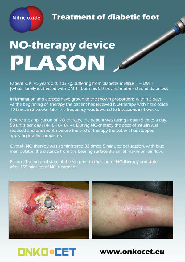
The PDF with the short report with pictures from the therapy of a diabetic foot can be viewed or downloaded here.
The pictures from the treatment of unhealing wounds an be found here:
http://www.onkocet.eu/en/produkty-detail/220/1/
The pictures from the treatment of unhealing wounds an be found here:
http://www.onkocet.eu/en/produkty-detail/293/1/
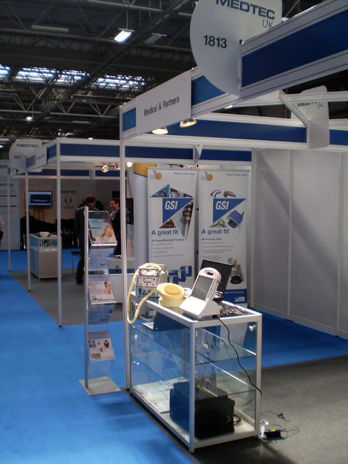
ONKOCET Ltd. has exhibited the devices from its portfolio on the MEDTEC UK exhibition in Birmingham, April 2011 through our partner Medical & Partners.
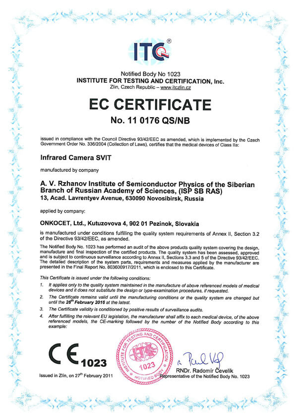 The ONKOCET company has successfully reached the certification of yet another medical device, Infrared Camera SVIT. The Certificate can be found here. The videos from the device operation can be found here.
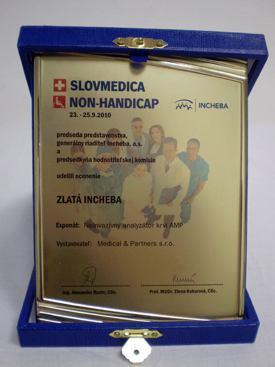 Our device, the non-invasive blood analyzer AMP has won the Golden Incheba prize at a medical exhibition SLOVMEDICA - NON-HANDICAP 2010. A big thank you goes to the organizers of the exhibition for acknowledging the quality of our device and to the exhibitor, the Medical & Partners company, for introduction of the AMP device to the medical public again.
We are pleased to inform our business partners, that our company has succesfully finished the certification process of Concor Soft Contact Lenses.
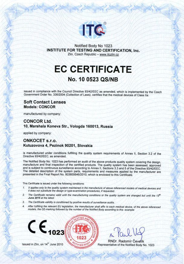 You can find the certificate here.
More information on Concor Soft Contact Lenses go to section Medical preparations/Concor soft contact lenses, or follow this link.
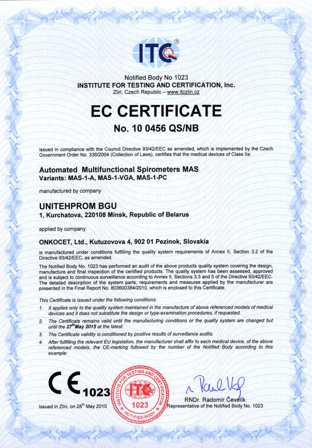 Our company has finished the certification process for another medical device, computerized spirometer MAS-1K with oximeter. You can find the device certificate here.
.jpg) Since May 2010 there is a new version of AMP device available.
Since May 2010 there is a new version of AMP device available.Follow this link if you want to see the pictures and specifications of the device.
http://www.onkocet.eu/en/produkty-detail/293/1/
 Dear partners,
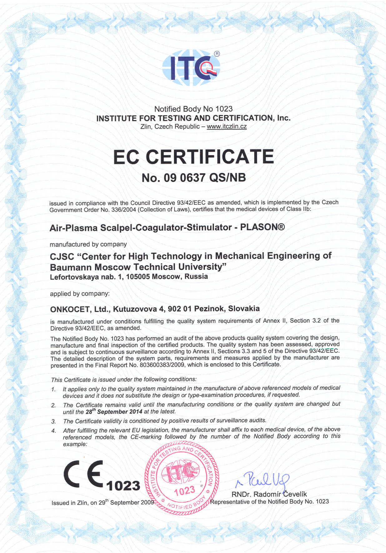
 In October 2009 we have received CE certificate for another device from our portfolio, NO therapeutical device PLASON. You can find more information about this revolutionary device, used for healing of unhealing wounds, diabetic foot, or for cosmetical purposes, at our webpage, section "Medical devices" -> PLASON-NO Therapy.
.gif)
Best regards
Team of ONKOCET Ltd. company
Post-Clinical Trial: Arkansas, USA
Radiothermography for detection of breast cancer by patients with palpable or mammographically detectable breast masses
Jacobo Nurko Ma.D, Department of Surgery,
University of Arkansas for Medical Sciences
Anne T. Mancino M.D.
Associate Professor, Section of Surgical Oncology and General Surgery, Department of Surgery
J. Ralph Broadwater, M.D.
Associate Professor and Section Chief, Section of Surgical Oncology
Bonny H.Wallace, MHSA, CCRP.
Director of Clinical Research
Michael J. Edwards, M.D.
Professor and Chairman, Department of Surgery
University of Arkansas for Medical Sciences
4301 W. Markham, Slot 725
Little Rock, AR 72205
ABSTRACT
INTRODUCTION
Radiothermography (RTM) has shown diagnostic value in the detection of breast cancer in international studies, but this data has not been reproduced in the United States. We evaluated the diagnostic efficacy of the BreastScan RTM device for the detection of breast cancers in patients with suspicious breast lesions on physical exam or breast imaging who were scheduled for breast biopsy. We measured the specificity, sensitivity, accuracy, positive predictive value and negative predictive value of RTM by comparing preoperative RTM findings with the resulting histology from biopsy.
METHODS
Patients presenting to the Breast Oncology Clinic with mammographic findings indicating need for biopsy were screened for participation in the study. Forty three patients underwent RTM scan prior to biopsy; RTM data were sent to a blinded reviewer for interpretation. Suspicious RTM results were compared to mammogram to confirm concordance. Breast biopsies were performed based on initial mammogram abnormalities. Definitive pathology results from breast biopsy were used as the standard for statistical comparison with the results of the RTM to determine the diagnostic parameters of sensitivity, specificity, positive predictive value and negative predictive value.
RESULTS
The diagnostic parameters for RTM were as follows: sensitivity of 84%, specificity of 70%, positive predictive value of 68%, and a negative predictive value of 85%.
CONCLUSIONS
An early U.S. experience with RTM confirms the reported Russian experience. The diagnostic efficancy of radiothermography in the detection of breast cancer is similar to that of mammography. Because RTM uses a completely distinct physical entity to detect breast cancer there is the possibility that a combination of RTM and diagnostic mammography may significantly enhance our ability to detect breast cancer.
There will be over 200,000 new cases of breast cancer in the U.S. this year and almost 40,000 patients will succumb to this disease.
Breast cancer is the most common malignancy in women and accounts for one-third of all new cancers in women. Mammography, breast self-exam and physical exam are the primary modalities for detecting breast cancer. Detected suspicious lesions are biopsied and examined pathologically for the presence of malignancy. The diagnostic efficacy of mammography and ultrasound to identify breast lesions as malignant has been well established in numerous studies. The sensitivity is generally accepted as 80 to 90 % and varies depending on breast density. Sensitivity also improves with advancing age. Specificity is general regarded in the >90% range and also depends on breast density.
The limitations of our currently available screening and diagnostic technologies have resulted in a large number of women being subjected to surgical and image-guided breast biopsy for definitive diagnosis.
Estimates are that approximately 1.8 million women undergo diagnostic breast biopsy annually in North America. It would be obviously beneficial if non-invasive diagnostic techniques were available to improve the diagnostic efficacy of our current screening and diagnostic breast evaluations and minimize the number of invasive and more costly procedures.
RTM has been tested in three international clinical studies to date. In one study, involving 81 patients, 48 were found to have breast cancer (8). RTM had a sensitivity of 90%, an accuracy of 86% and a specificity of 82%. In a second study involving 43 patients, 35 of which had breast cancer, RTM had a sensitivity of 94%, an accuracy of 90% and a specificity of 78%. In the largest study, involving 771 patients, of which 101 were found to have breast cancer, RTM had a sensitivity of 85%, an accuracy of 78% and a specificity of 77% .
While this early experience with the technology is remarkably encouraging, no series of patients has to date been evaluated in North America. Based on the findings of early studies with RTM in the detection of breast cancer, we designed this study for proof of concept testing in North America to more accurately estimate its diagnostic accuracy.
METHODS
The objective of this study was to measure the specificity, sensitivity, accuracy, positive predictive value and negative predictive value of radiothermography in the diagnosis of breast cancer in women with palpable or mammographically evident breast masses who are undergoing breast biopsy.
Collected RTM data was analyzed in conjunction with the pathology report of the breast biopsy to determine parameters of the diagnostic efficacy (sensitivity, specificity, positive predictive value, negative predictive value and accuracy) of the BreastScan device.
Patients presenting to the Breast Oncology Clinic with mammographic findings indicating need for biopsy were screened for participation in the study. Forty three patients underwent RTM scan prior to biopsy; RTM data were sent to blinded reviewer for interpretation. Suspicious RTM results were compared to mammogram to confirm concordance. Breast biopsies were performed based on initial mammogram abnormalities. Definitive pathology results from breast biopsy were used as the standard for statistical comparison with the results of the RTM to determine the diagnostic parameters of sensitivity, specificity, positive predictive value and negative predictive value.
All enrolled patients met the following criteria to be eligible for the study:
1) minimum age of 30,
2) found to have an abnormal mammogram either BIRADS IV or V, (Table I) alone or in combination with abnormal physical exam,
3) prior to enrollment all were scheduled for a diagnostic breast biopsy to be performed as an “open” surgical breast biopsy or by a needle core tissue biopsy with a 16 or larger gauge needle,
4) all provided written informed consent and signed a research authorization for access to protected health information,
5) for pre-menopausal patients, the collection of RTM data must have occurred within 6 – 9 days after the end of the patients last menstrual day.
Patients were excluded from the study if they were found to have:
1) pregnancy or lactation within 6 months of study entry,
2) a prior history of a percutaneous or surgical breast procedure within 6 months of study,
3) a prior history of breast augmentation, reduction mammoplasty, or reconstruction,
4) a prior history of breast radiotherapy,
5) a prior history of a breast ductal cannulation within six months of study enrollment,
6) a prior history of a medical condition, (including mental illness) that in the opinion of the investigator might potentially interfere with the patient’s participation or compliance with the evaluation.
Table I- BIRADS Classification.
Technology
The Breast Scan is a medical electromagnetic radiometer for the percutaneous, noninvasive temperature assessment of internal structures. The device consists of two antennas/scanners (Infrared and Microwave), which detects thermal abnormalities at a depth of between 3 – 7 cm with an accuracy of + 0.2 o for temperatures of 32 – 38 o C. The antenna contacts the skin and detects naturally produced electromagnetic radiation proportional to temperature. Tissue emits electromagnetic radiation. This relationship is described by the equation:
P = K x T x B
where:
P is the power received in Watts
K is Boltzmann’s constant
B is the frequency (in Hz) and
T is the temperature
Because a known temperature produces a known power, measuring the power received determines the temperature at a given frequency of radiation. The radiation signal is picked up from the breast by the Breast Scan’s antennas and is amplified in the internal temperature sensor; the signal is then transmitted to the data processing unit, which can then be transferred to a personal computer, where it is stored and where the temperature data can be integrated and graphed into an isotherm image to show temperature differentials at corresponding points for each breast. This method allows analysis of temperature differentials between the corresponding points, and thus able to identify asymmetries. One elegant feature of breast temperature measurements by this technique is that each woman serves as her own control, so that internal temperature disparities (if they exist) are immediately recognized.
Unlike standard thermography, which measures temperature at the skin, RTM senses and measures the temperatures of deeper internal structures.
Another important feature of the software of the Breast Scan technology is that temperature data are also displayed as a temperature field with isotherm lines [Fig- 1] similar to that used for traditional infrared thermography. In the temperature field, cool areas of the breast are displayed by "cold" colours (i.e. blue) and hot ones are reflected by "warm" colours (red and orange). Internal temperature fields show temperature abnormalities, in particular, corresponding to the location of rapidly growing tissue. Similar fields are constructed with use of the infrared measurement device and displayed as temperature fields of surface skin temperatures (similar to Figure 1).
Skin temperatures are generally cooler than deep internal temperatures. In addition an overlay graph is plotted at individual temperature measurement sites. There are nine different measurement location sites for each breast, and measurements are also made of the axillary temperatures. When the internal microwave and infrared skin temperature measurements are overlaid on this plot, additional information is extrapolated when the skin and the deep internal temperature readings demonstrate similar thermal asymmetry at the same location [right versus left breast]. Asymmetries in skin temperatures alone are particularly predictive of cancer when women are thin and have extremely small breasts. Asymmetries in deep internal temperatures are predictive of cancer for women with normal size to very large breast.
In the example shown as Figure 1, the microwave temperature field of the right breast is shown on the left of the figure, and the temperature field of the left breast is shown on the right of the figure (as if you were looking at the patient in front of you). In this particular figure, the temperature field demonstrates a large thermal asymmetry of the left breast with the central temperature reading at the nipple 1.1?C warmer on the left than on the right. Biopsy results confirmed a malignancy in this location in the left breast.
Figure 1. Temperature fields of the right and left breast, with isotherm lines. This temperature field was constructed from deep internal temperature readings with the microwave receiver. The two blue temperature circles displayed in the central portion of the figure represent reference temperature readings of the pleural and abdominal cavities respectively. The two green temperature circles displayed in the upper outer portions of the figure represent temperature readings of the axillary lymph nodes.
Microwave radiometry detection of the “thermal signal” from rapidly growing tissue is based upon measurement of the intensity of natural electromagnetic radiation from a patient’s tissue at microwave frequencies. This electromagnetic radiation intensity is directly proportional to the temperature of the tissue. Areas of thermal abnormality may be caused by higher metabolic activity of tumors, and this higher metabolic activity may be generated from the angiogenesis of the tumor or the immune inflammatory response to the tumor. According to M. Gautherie, (1) temperature and blood flow pattern in cancerous mammary tissues result from two phenomena: heat transfer from the cancer into the surrounding tissues and vascular reactions. The relative contribution of heat depends on the actual vascularization, which is largely different from one breast to the other, partic¬ularly under malignant conditions. The metabolic heat produced by the tumor is transferred to the surroundings tissues, particularly towards the skin so that increases in skin temperature are generally associated with cancer.
RESULTS:
To date 43 evaluable patients have been enrolled and completed the study at the University of Arkansas for Medical Sciences Campus at this time; however data from 9 examinations were deleted from the statistics due to “criteria” deviations [Table II]. A mammogram on the same day of the examination causes excessive manipulation of breast tissue with disruption of normal thermal gradients. On several occasions it was not known at the time of the examination that a woman had undergone a mammographic examination the same day. This information was obtained in retrospect. We also learned that an ultrasound procedure also disrupts the normal thermal gradients of breast tissue. These questions are now asked as part of the inclusion/exclusion criteria questions during screening.
There were 2 indeterminate tests that do not fit into the above classifications: 1 was malignant and 1 was benign. These 2 exams are not considered in the statistical analysis below. 2 additional patients are awaiting biopsy at this time.
From the 30 patients considered in the statistical analysis, there were 11 true positive cases, 5 false positive, 2 false negative, and 12 true negative cases providing a sensitivity of 84% and a specificity of 70% with a negative predicted value of 85% and a positive predicted value of 68%. The negative and positive likelihood ratios are 0.21 and 2.8 respectively. (Table III).
True Positive, n=2; True Negative, n=1; False Positive, n=2; False Negative, n=4;
(A) True Positives = 11
(B) False Positives = 5
(C) False Negatives = 2
(D) True Negatives = 12
? (Total) 30
Table III. RTM BreastScan Accuracy Report of 30 Patients
The following accuracy statistics are calculated based on variables A, B, C, and D:
Sensitivity: A/A+C = 84%
Specificity: D/B+D =70%
Negative Predictive Value = D/C+D = 85%
Positive Predictive Value = A/A+B = 68%
Negative Likelihood Ratio = C/A+C ÷ D/B+D = 0.21
Positive Likelihood Ratio = A/A+C ÷ B/B+D = 2.8
DISCUSSION:
Mammography and physical exam are the current tools most commonly used for the detection of breast cancer, unfortunately approximately 15% of breast cancers cannot be imaged with mammogram, ultrasound or MRI, which is particularly true in young women with dense breasts and for women with fibrotic or fibrocystic changes needing a different approach. These varieties of methods available for clinical exploration of the breast provide information essentially on morphology and structure. By contrast, the thermal methods, among them RTM using infrared and microwave scanners, offer the advantage of giving information on thermophysiology, the superficial and internal thermal pattern of the breast in relation to the metabolism and vascularization within its underlying tissue. Under these circumstances, we can speculate that RTM, in conjunction with mammography can provide an applicable role in the accurate and early diagnosis of breast cancer in 98 to 99% of the cases. Anecdotally, in a courtesy examination for a non-protocol patient with a breast palpable mass, RTM was able to identify breast cancer confirmed as lobular cancer by histology that was not detected by mammography, ultrasound nor MRI.
The essence of breast thermography lies in the detection of differences in the bilateral thermal symmetry, corresponding to a local or general increase in temperature level and/or change in thermal pattern. (2).
Until the middle of the 1950’s infrared thermography was used almost exclusively as a military tool, but it has since become available for civilians usage. In medicine the greatest interest was given to the diagnosis of breast cancer. The first paper concerning this matter was published in 1956 by Lawson (3) who found an increased temperature of the skin over some breast cancers. After Lawson’s original paper, many papers have been published about thermography and diagnosis of breast cancer that have shown that increased intratumoral and peritumoral temperature, as a consequence of an increase on blood flow and metabolic rates, is a key factor which can be targeted to precisely localize a breast lesion. However several other authors suggested that thermography was not a sufficiently precise modality for use in routine breast diagnosis and also there was a clear need to develop an objective system for evaluation of breast thermograms, since none of the systems available for evaluation of breast thermograms were used without criticism (10-12) . This controversy of thermography as a tool for diagnosis of breast cancer took place in the late 1960’s and 70’s where no consideration of possible variability in temperature caused by multiple factors was made appropriately. Currently it is well known that several factors like circadian rhythm, breast size, body habitus, menstrual cycle, pregnancy, hormonal replacement therapy, manipulation of the breast prior to thermographic study, recent interventional breast procedures, and some others factors that were unknown or under investigation back in those times, can modify the thermal pattern of the breast and simultaneously provide erroneous results on thermogram interpretations. Another drawback that simple thermography (infrared and/or liquid crystal) faced in the past, was its inability to measure deeper breast temperatures supporting their thermographic findings only from skin or superficial thermal patterns. These aspects mentioned above, were the reasons we believe simple thermography never obtained the precision expected losing credibility and being abandoned as a tool in the screening or diagnosis of breast cancer.
Gautherie (1,4,6,7), in 1980 thinking ahead and in his efforts to demonstrate that simple thermography is a potent tool in the aid of diagnosis of breast cancer, knew that measuring deeper temperatures within the breast tissue might add important value to already promising results, thus he implanted intramammary probes (6.5cm fine needles) in the cancerous breast as well as the contralateral healthy breast in approximately symmetrical locations. In the cancerous breast, a radiography test was made in order to check that the needle was passing through the tumor close at its center. He demonstrated that thermal conductivity of cancer is twice as high as the healthy tissue. Although Gautherie added an important value to thermography he did not gain acceptance due to the invasiveness and complexity of the procedure.
RTM on the other hand, has the ability to measure superficial temperatures with an infrared scanner as well as the capacity to measure internal or deeper thermal patterns with the aid of a microwave scanner in an non-invasive manner; therefore providing a complete distribution of temperatures in the breast bilaterally that are analyzed by a computerized system which integrates and displays the thermal patterns of both breasts, and compares one to another searching for temperature differences and patterns.
This study, with the aid of RTM, clearly demonstrated that the presence of both hyperthermia and hypervascularization within the tumor, as well as at its periphery in relation to the temperature and blood flow in the contralateral healthy breast tissue, are dominant signs for detection of a breast lesion.
Herein, we present our initial experience in a small, but well-defined, group of patients, all of whom had a breast radiothermographic evaluation done at the same time as the establishment of a definitive diagnosis by breast biopsy. The preliminary result clearly shows promising results that RTM for the detection of breast cancer in patients with a palpable or mammographically detectable breast mass may contribute to the detection of breast cancer, and the identification of women at high risk of developing breast cancer.
In our experience, RTM has shown a sensitivity of 84%, and a specificity of 70%. Interestingly, Of the 30 patients that were identified as probably having breast cancer by mammogram as BIRADS IV or V (table I), we found that RTM identified 11 out of the 13 cases with diagnosed breast cancer by biopsy, meaning that the number of biopsies performed can be reduced if we rely on RTM.
All five false positives cases (table.IV) were histologically confirmed as proliferative lesions; RTM can reasonably be expected to have a higher false positive rate than mammography since it is able to localize benign processes or pre-malignant lesions while they are at early stages; this is most probably related to a higher metabolic phase or angiogenesis; whether RTM will enhance our capacity to identify patients at increase risk for developing breast cancer is not yet known, but is clearly a potential benefit to consider.
Gautherie, et al., (1,4,6,7) in their efforts to implement thermography as a useful screening method for breast cancer, published a series of more than 25,000 cases where thermography incorrectly diagnosed breast cancer in 958 patients that did not have it based on physical exam or mammographic examinations, but later in their long term follow-up (length interval of 4-41 months), 204 (21%) patients developed breast cancer. Another report from Wallace. JD, et al., (5) also published a series of 597 patients that underwent thermography, physical exam and mammography. The thermographic exam was positive, but the mammographic exams of 11 cases were interpreted as negative, and the opinion of the referring physician on clinical evidence did not warrant biopsy. Subsequently, between 2 to 10 months, all these patients developed clinical or mammographic signs of cancer with histological confirmation.
Combined RTM and mammography enhance our diagnostic efficacy and are complementary in enhancing our sensitivity to detect breast cancer.
RTM also has potential future usefulness as a screening method for breast cancers. Longer term follow-up data is needed however, particularly as it pertains to RTM abnormalities in areas where no mammographic lesion is seen.
It is clear that we are in too early a stage of development to build definitive conclusions regarding the actual role of RTM and avoid innocent acceptance of this new technology that can result in misuse of a diagnostic modality; but it is also true that breast RTM appears to have an important value based on our preliminary results.
BIBLIOGRAPHY REFERENCES:
1. Gautherie, M. Temperature and blood flow patterns in breast cancer during natural evolution and following radiotherapy, Prog Clin Biol Res. 1982; 107:21-64
2. Johansson, N.T. Thermography of breast. A clinical study with special reference to breast cancer detection. Acta Chir Scand Suppl. 1976; 460:3-91
3. Lawson, R.N. Implications of surface temperatures in the diagnosis of breast cancer, Can Med Assoc J. 1965; 75: 309-10.
4. Gautherie, M, Haehnel P, Walter J.P. et al; Thermovascular changes associated with in situ and minimal breast cancer. Results of an ongoing prospective study after four years. J Reprod Med. 1987 Nov; 32(11):833-42.
5. Wallace, J.D. Thermography in the diagnosis of breast cancer. Radiology. 1968 Oct;91 (4):679-85
6. Gautherie, M. Thermopathology of breast cancer: measurement and analysis on in vivo temperature and blow flow. Ann N Y Acad Sci. 1980; 335:383-415.
7. Gautherie, M. Thermobiological assessment of benign and malignant breast disease. Am J Obstet Gynecol. 1983 Dec 15;147(8);861-9.
8. Sdvigkov AM, Vesnin SG, Kartashova AF, Bjahov MJ, Gurtovoi IJ, Borisov VI, Arablinskii VM, Kozlovskii OM, Sobol MJ, Goncharov BJ, Torlina VE, Makarova EE, Popova SV. On the Place of Radio-Thermometry in Mammological Practice. Aktualnyie Problemy Mammalogii, pp 28-40, 2000.
9. Libshitz H,I. Thermography of the breast. Current status and future expectations. JAMA. 1977 Oct 31; 238(81):1953-4.
10. Sterns E.E, Zee B. Thermography as a predictor of prognosis in cancer of the breast, Cancer. 1991 Mar 15;67(6):1678-80.
11. Sterns E.E, Curtis, A.C, Miller, S. et al; Thermography in breast diagnosis. Cancer. 1982 Jul 15;50(2):323-5
12. Isard, H.J. Sweitzer, C.J. Edelstein, G.R. Breast thermography. A prognostic indicator for breast cancer survival. Cancer. 1988 Aug 1;62(3):484-8.

