Medical devices
search
news
Pictures from NO therapy of diabetic foot by PLASON device 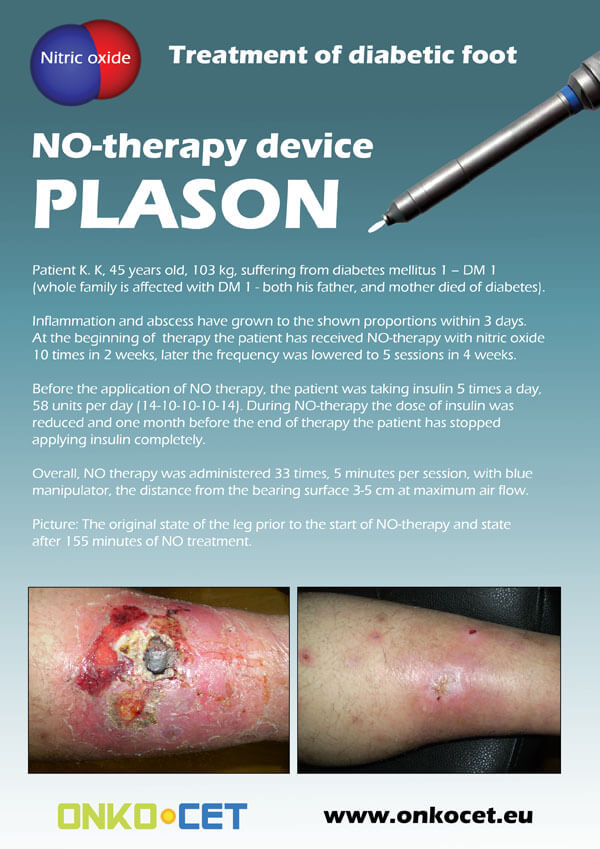
The PDF with the short report with pictures from the therapy of a diabetic foot can be viewed or downloaded here.
The pictures from the treatment of unhealing wounds an be found here:
http://www.onkocet.eu/en/produkty-detail/220/1/
The pictures from the treatment of unhealing wounds an be found here:
http://www.onkocet.eu/en/produkty-detail/293/1/
12.5.2011 12:23
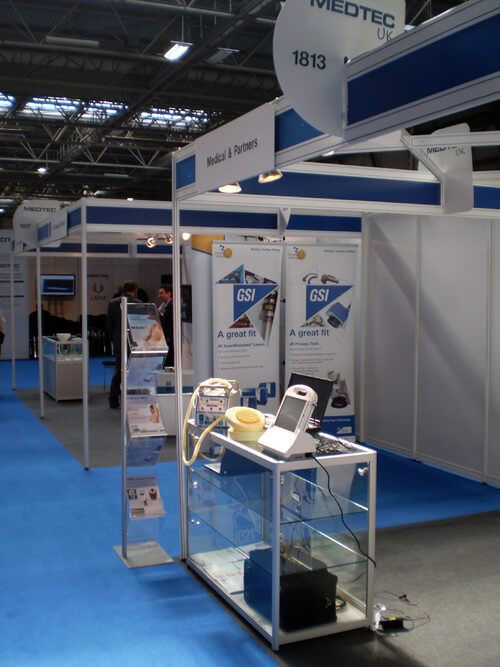
We have exhibited our devices on the prestigious MEDTEC UK exhibition in Birmingham
ONKOCET Ltd. has exhibited the devices from its portfolio on the MEDTEC UK exhibition in Birmingham, April 2011 through our partner Medical & Partners.
12.4.2011 12:27
Certification of SVIT infrared camera completed
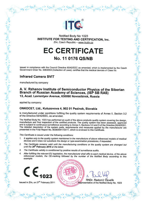 The ONKOCET company has successfully reached the certification of yet another medical device, Infrared Camera SVIT. The Certificate can be found here. The videos from the device operation can be found here.
 The ONKOCET company has successfully reached the certification of yet another medical device, Infrared Camera SVIT. The Certificate can be found here. The videos from the device operation can be found here.
 4.3.2011 11:38
Golden Incheba prize for AMP device
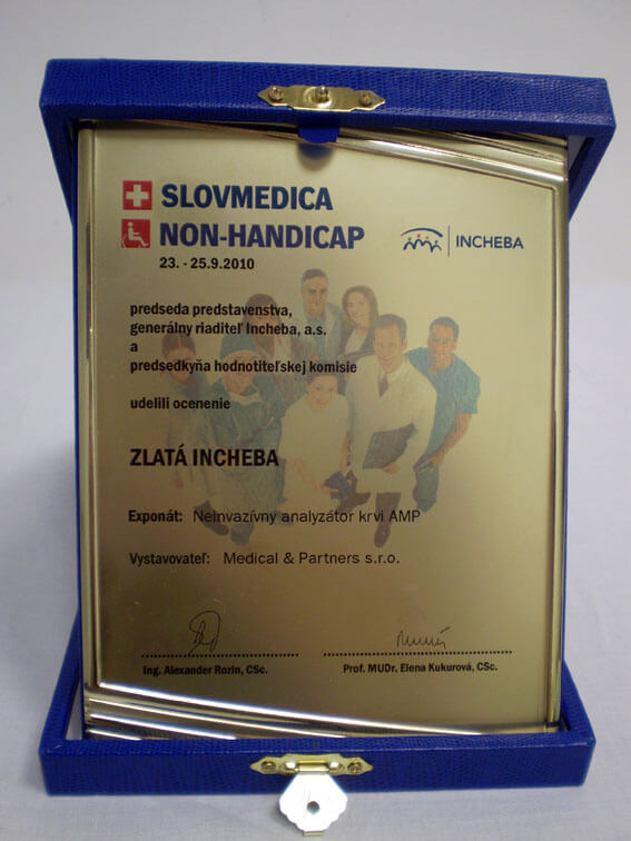 Our device, the non-invasive blood analyzer AMP has won the Golden Incheba prize at a medical exhibition SLOVMEDICA - NON-HANDICAP 2010. A big thank you goes to the organizers of the exhibition for acknowledging the quality of our device and to the exhibitor, the Medical & Partners company, for introduction of the AMP device to the medical public again.
 Our device, the non-invasive blood analyzer AMP has won the Golden Incheba prize at a medical exhibition SLOVMEDICA - NON-HANDICAP 2010. A big thank you goes to the organizers of the exhibition for acknowledging the quality of our device and to the exhibitor, the Medical & Partners company, for introduction of the AMP device to the medical public again.
 4.10.2010 11:29
Certification of Concor Soft Contact Lenses successfully finished.
We are pleased to inform our business partners, that our company has succesfully finished the certification process of Concor Soft Contact Lenses.
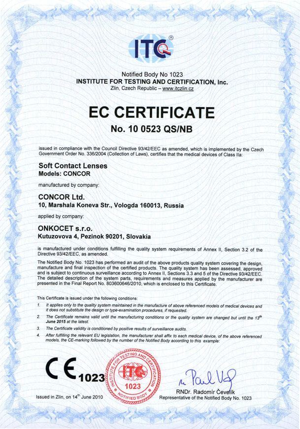 You can find the certificate here.
More information on Concor Soft Contact Lenses go to section Medical preparations/Concor soft contact lenses, or follow this link.
We are pleased to inform our business partners, that our company has succesfully finished the certification process of Concor Soft Contact Lenses.
 You can find the certificate here.
More information on Concor Soft Contact Lenses go to section Medical preparations/Concor soft contact lenses, or follow this link.
16.6.2010 11:51
Certification of MAS-1K Spirometer successfully finished.
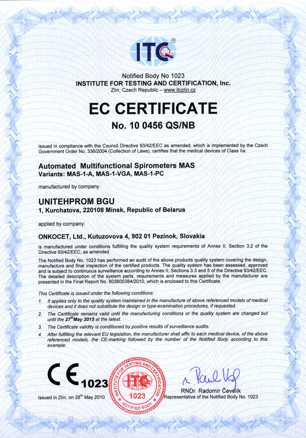 Our company has finished the certification process for another medical device, computerized spirometer MAS-1K with oximeter. You can find the device certificate here.
 Our company has finished the certification process for another medical device, computerized spirometer MAS-1K with oximeter. You can find the device certificate here.
 2.6.2010 10:58
New version of AMP device available
.jpg) Since May 2010 there is a new version of AMP device available.
Since May 2010 there is a new version of AMP device available.
Follow this link if you want to see the pictures and specifications of the device.
.jpg) Since May 2010 there is a new version of AMP device available.
Since May 2010 there is a new version of AMP device available.Follow this link if you want to see the pictures and specifications of the device.
11.5.2010 15:17
Treatment of burns with PLASON We added photographs of treatment of burns with PLASON device:
http://www.onkocet.eu/en/produkty-detail/293/1/
http://www.onkocet.eu/en/produkty-detail/293/1/
17.3.2010 12:08
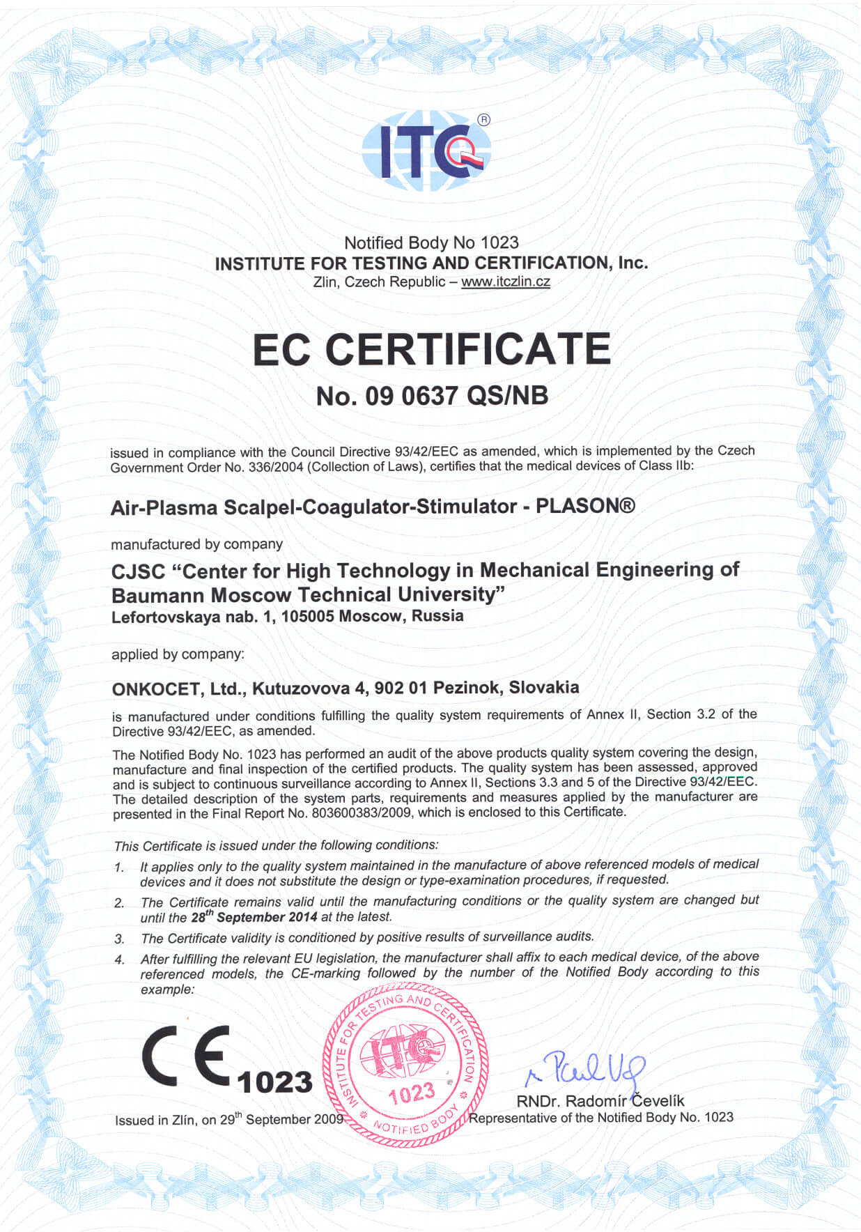
Certification of PLASON device finished, CE issued.
 Dear partners,
In October 2009 we have received CE certificate for another device from our portfolio, NO therapeutical device PLASON. You can find more information about this revolutionary device, used for healing of unhealing wounds, diabetic foot, or for cosmetical purposes, at our webpage, section "Medical devices" -> PLASON-NO Therapy.
.gif)
Best regards
Team of ONKOCET Ltd. company
 Dear partners,
 In October 2009 we have received CE certificate for another device from our portfolio, NO therapeutical device PLASON. You can find more information about this revolutionary device, used for healing of unhealing wounds, diabetic foot, or for cosmetical purposes, at our webpage, section "Medical devices" -> PLASON-NO Therapy.
.gif)
Best regards
Team of ONKOCET Ltd. company
23.1.2009 15:30
Introduction
Sonomed-500
| |||||||||||||||||||||||
| |||||||||||||||||||||||
  B+D B+PD Sonomed-500 combines the following display modes: B, B+B, M, B+M, D, B+D, B+CFM, B+CFM+D, B+PD, B+PD+D. They enable to obtain the visualization of abdominal organs as well as carry out echocardiographic or vascular examinations. Current settings of the system can be saved for routine examinations. The four focusing zones and the possibility of their selective switching on ensure an optimal quality of the image in any mode. | |||||||||||||||||||||||
| |||||||||||||||||||||||
| |||||||||||||||||||||||
| |||||||||||||||||||||||
| Specialized programmes | |||||||||||||||||||||||
| |||||||||||||||||||||||
| Comprehensive database | |||||||||||||||||||||||
| |||||||||||||||||||||||
The software package ensures:
- support of several databases;
- work with several databases through a network;
- generating report on the base of templates.
Searching facilities make the database easy to use when working with large-scale data.
The graphic interface of the database gives maximum comfort and saves your time when working with images and reports from the archive. Reports are based on ready-made templates.
